Amino Acid 3D Explorer
All 20 amino acids as rotatable 3D models, grouped by side chain. Drag the pH and watch each charge change, with every pKa and pI marked.
Visualiser
Tap a card to open it in 3D, or move between cards with the arrow keys. Space plays and pauses.
At pH 7.40, 4 carry half a charge or more: lysine +0.97, arginine +0.98, aspartic acid −1.01, glutamic acid −1.00. Partly charged: cysteine −0.14. The other 15 are within 0.05 of zero.
Histidine His · H Positively charged
- Net charge at this pH The sum over its ionisable groups, each by Henderson-Hasselbalch: α-carboxyl −1.00, α-amino +0.98, imidazole +0.04.
- +0.02
- Dominant form The commonest protonation state and its share of the molecules: the product of each group’s share, since the groups are treated as independent.
- zwitterion 95%
- pI The mean of the two pKas either side of the neutral form, which is also where the model’s net charge is zero.
- 7.59
- pK₁ (α-COOH) Nelson and Cox, Lehninger Principles of Biochemistry, 7th edition, Table 3-1. Below this pH most of the α-carboxyl groups carry their proton.
- 1.82
- pK₂ (α-NH₃⁺) Nelson and Cox, Lehninger Principles of Biochemistry, 7th edition, Table 3-1. Below this pH most of the α-amino groups carry the third proton.
- 9.17
- pKR (side chain) The imidazole group, a base: it carries a proton below this pH. Nelson and Cox, Lehninger Principles of Biochemistry, 7th edition, Table 3-1.
- 6.00
- Side chain The R group’s functional group. Positively charged, in Lehninger’s five classes, as the Protein Synthesis Simulator colours them; some books move glycine, cysteine, histidine or tyrosine.
- imidazole
- Formula The uncharged molecule, NH₂ and COOH. The zwitterion has the same atoms.
- C₆H₉N₃O₂
- Molar mass From the formula and the IUPAC standard atomic weights the site uses everywhere.
- 155.16 g/mol
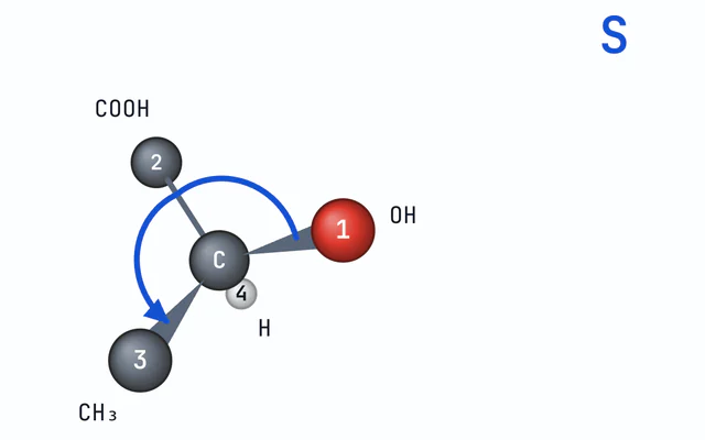
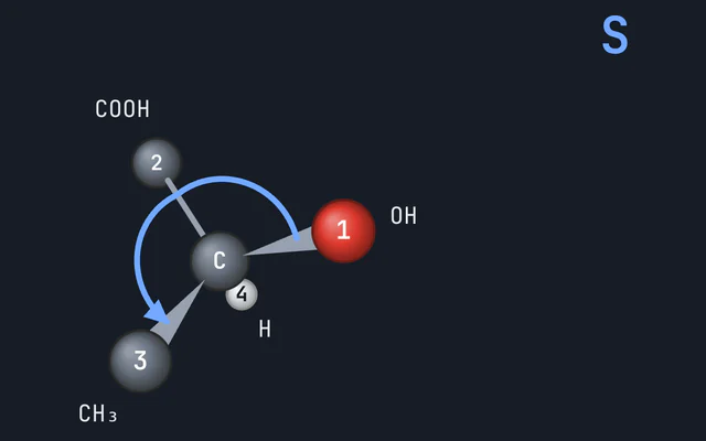
- Configuration at Cα L, as in every protein, and S by the Cahn-Ingold-Prelog rules: NH₂ outranks COOH, which outranks the side chain, with H last.
- L, S
- Hydropathy Kyte and Doolittle (1982): from isoleucine at 4.5, the most hydrophobic, to arginine at −4.5.
- −3.20
- Codons The mRNA codons for histidine in the standard genetic code, 2 of them.
- CAU CAC
- pKa values
- Net positive
- Net negative
- pI 7.59
Citing this tool
Last updated . Add the date you accessed it as well, which a citation of a page that can change asks for. If a specific result matters, cite the permalink from the tool’s share row instead of this page: it reproduces the exact parameters.
On the syllabus: AP Biology, Unit 1: Chemistry of Life; MCAT, 1A: Structure and function of proteins and their constituent amino acids.
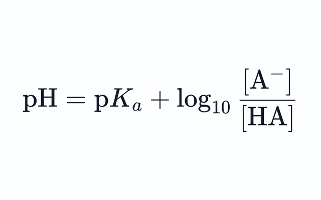
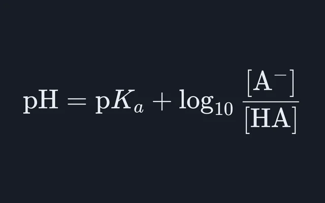
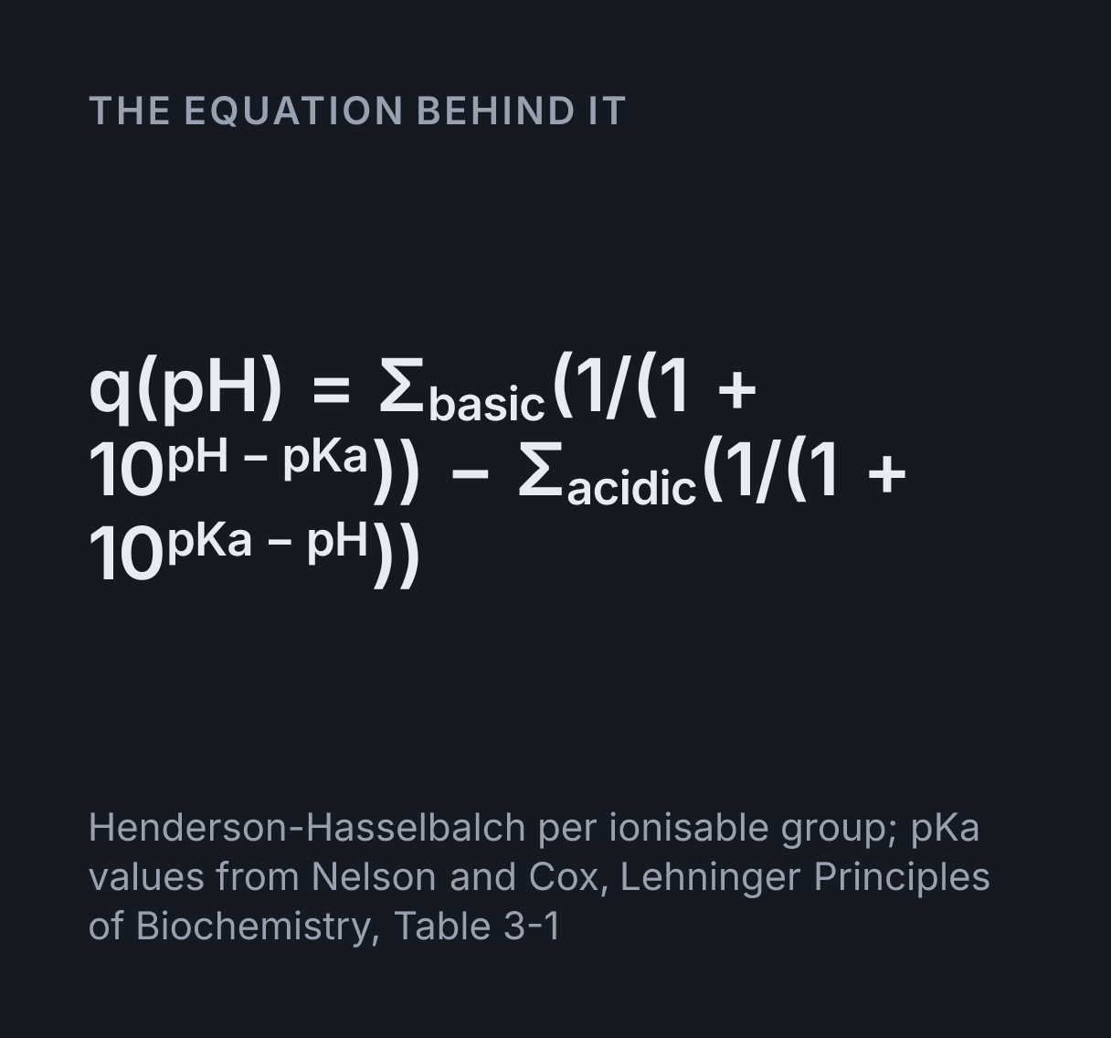
The equation
Henderson-Hasselbalch per ionisable group; pKa values from Nelson and Cox, Lehninger Principles of Biochemistry, Table 3-1
What are the 20 standard amino acids and their functional groups?
The 20 standard amino acids share a core backbone: a central alpha-carbon carrying an amino group (−NH2, or in proline a ring −NH−), a carboxyl group (−COOH), a hydrogen atom and a distinguishing side chain (R group). They are categorised into non-polar aliphatic, aromatic, polar uncharged, positively charged (basic: Lys, Arg, His), and negatively charged (acidic: Asp, Glu) residues.
The 20 amino acids and their side chains
The side chain is the only part that differs, and it decides the class. Lehninger’s textbook sorts the 20 into five classes: 7 nonpolar and aliphatic, 3 aromatic, 5 polar and uncharged, 3 positively charged and 2 negatively charged. The chart opens in that order, each card topped with its class colour.
| Amino acid | Codes | Class | pK₁ | pK₂ | pKR | pI | Charge at pH 7.4 |
|---|---|---|---|---|---|---|---|
| Glycine | Gly, G | Nonpolar, aliphatic | 2.34 | 9.60 | none | 5.97 | −0.01 |
| Alanine | Ala, A | Nonpolar, aliphatic | 2.34 | 9.69 | none | 6.01 | −0.01 |
| Proline | Pro, P | Nonpolar, aliphatic | 1.99 | 10.96 | none | 6.48 | 0.00 |
| Valine | Val, V | Nonpolar, aliphatic | 2.32 | 9.62 | none | 5.97 | −0.01 |
| Leucine | Leu, L | Nonpolar, aliphatic | 2.36 | 9.60 | none | 5.98 | −0.01 |
| Isoleucine | Ile, I | Nonpolar, aliphatic | 2.36 | 9.68 | none | 6.02 | −0.01 |
| Methionine | Met, M | Nonpolar, aliphatic | 2.28 | 9.21 | none | 5.74 | −0.02 |
| Phenylalanine | Phe, F | Aromatic | 1.83 | 9.13 | none | 5.48 | −0.02 |
| Tyrosine | Tyr, Y | Aromatic | 2.20 | 9.11 | 10.07 | 5.66 | −0.02 |
| Tryptophan | Trp, W | Aromatic | 2.38 | 9.39 | none | 5.89 | −0.01 |
| Serine | Ser, S | Polar, uncharged | 2.21 | 9.15 | none | 5.68 | −0.02 |
| Threonine | Thr, T | Polar, uncharged | 2.11 | 9.62 | none | 5.87 | −0.01 |
| Cysteine | Cys, C | Polar, uncharged | 1.96 | 10.28 | 8.18 | 5.07 | −0.14 |
| Asparagine | Asn, N | Polar, uncharged | 2.02 | 8.80 | none | 5.41 | −0.04 |
| Glutamine | Gln, Q | Polar, uncharged | 2.17 | 9.13 | none | 5.65 | −0.02 |
| Lysine | Lys, K | Positively charged | 2.18 | 8.95 | 10.53 | 9.74 | +0.97 |
| Arginine | Arg, R | Positively charged | 2.17 | 9.04 | 12.48 | 10.76 | +0.98 |
| Histidine | His, H | Positively charged | 1.82 | 9.17 | 6.00 | 7.59 | +0.02 |
| Aspartic acid | Asp, D | Negatively charged | 1.88 | 9.60 | 3.65 | 2.77 | −1.01 |
| Glutamic acid | Glu, E | Negatively charged | 2.19 | 9.67 | 4.25 | 3.22 | −1.00 |
pK₁ belongs to the α-carboxyl group, pK₂ to the α-amino group and pKR to the side chain, from Nelson and Cox, Lehninger Principles of Biochemistry, 7th edition, Table 3-1. Each charge is the explorer’s own, computed group by group.
Each amino acid has a page of its own, with its charge from pH 1 to 13 and its pI worked out: see the amino acid pKa and pI table.
Using the explorer
- The chart shows all 20 in 3D at one scale, so tryptophan is as much bigger than glycine as it really is. It opens with a sweep of the pH from 7.4 down to 1, up to 13 and back, and each card’s badge changes as its groups gain and lose protons. Press Sweep pH to run it again. With reduced motion set, it opens still, at pH 7.4.
- The pH slider, beside Play and Sweep pH, sets the pH for everything at once. The marks under it are blood plasma’s 7.4 and the pKa values and pI of the amino acid the readouts describe, and its track is blue where that amino acid is net positive and red where it is net negative, as the plot is.
- Tap a card, or choose from the Amino acid menu, to open it. Drag to turn it, and tap an atom to name it. A proton that comes and goes is drawn as faint as the share of molecules that have it, and each charged group glows blue for positive and red for negative, as strongly as it is charged.
- Hydrogens, Labels, Space-filling and D form, under an open amino acid, change how it is drawn, and the Plot menu switches the plot between net charge against pH and the titration curve. On a wide screen the stage also adds up its charge group by group, beside the molecule.
- Save image saves the chart or the molecule as it is, with the pH in the picture.
Worked example: histidine from pH 1 to 13
Histidine has three groups that ionise: its α-carboxyl, pK₁ = 1.82, its imidazole side chain, pKR = 6.00, and its α-amino group, pK₂ = 9.17. For each, the Henderson-Hasselbalch equation gives the share of molecules in which it carries its proton. At pH 7.4:
- α-carboxyl: deprotonated in
1/(1 + 10^(1.82 − 7.4))of molecules, which is 1.0000 to four places, so it contributes −1.0000. - α-amino: protonated in
1/(1 + 10^(7.4 − 9.17)) = 0.9833, contributing +0.9833. - Imidazole: protonated in
1/(1 + 10^(7.4 − 6.00)) = 1/(1 + 25.1) = 0.0383, contributing +0.0383. - Net charge:
0.9833 + 0.0383 − 1.0000 = +0.0216, which the badge rounds to +0.02.
Sweep the pH and the same sum gives +1.87 at pH 1, where most molecules carry both ring protons and the
carboxyl proton, +1.00 at pH 4, +0.50 at pH 6.00, where half the side chains are protonated, 0.00 at the
pI, 7.59, −0.50 at pH 9.17 and −1.00 at pH 13. The pI is the mean of the two pKas either side of the
neutral form, (6.00 + 9.17)/2 ≈ 7.59.
The isoelectric point and the tyrosine exception
The pI is the pH at which an amino acid carries no net charge, so it does not move in an electric field.
For an amino acid with two ionisable groups it is the mean of their pKas: glycine’s is
(2.34 + 9.60)/2 = 5.97. With a third group, take the two pKas either side of the neutral
form. Aspartic acid’s neutral form lies between its two carboxyls, so its pI is
(1.88 + 3.65)/2 ≈ 2.77; lysine’s lies between its two amino groups, so its pI is
(8.95 + 10.53)/2 = 9.74.
The explorer also finds, by bisection, the pH where its own net charge curve crosses zero. For 19 amino acids that agrees with the table to within 0.01. Tyrosine is the exception: 5.63 against the table’s 5.66, and here the table is right. A titration measures each pKa for the molecule as a whole, so tyrosine’s α-amino pKa, 9.11, already counts the few molecules whose phenol, pKR 10.07, gives up its proton first. The explorer treats each group on its own and adds the phenol on top: at pH 5.66 about 4 in 100,000 of its phenols are ionised, beside the 35 in 100,000 that have lost an α-amino proton, so it counts about a tenth too many negative molecules. The net charge curve is so flat there that this moves the zero crossing by 0.02. The pI readout gives the table’s 5.66, with the curve’s 5.63 beside it.
Titration curves
Switch the plot to the titration curve to see pH against the equivalents of OH⁻ added, starting from the fully protonated amino acid at pH 0, as MCAT courses draw it. Each pKa is a flat stretch, a buffer region, where the pH is the pKa with that group half titrated. Between two of them the curve climbs steeply, and the pI sits in the middle of that climb: at about 1.00 equivalent for glycine, of the 2.00 it takes to go from pH 0 to 14. An amino acid with an ionisable side chain takes about 3 equivalents and has three flat stretches.
For one group at a time, the Henderson-Hasselbalch calculator gives the ratio of acid to base at any pH, and the titration curve calculator draws the curve for a weak acid titrated with a strong base, volumes and all.
L, S and the cysteine exception
Every amino acid except glycine has four different groups on its α-carbon, so it has two mirror-image forms, L and D, and proteins are built from L amino acids only. Press D form under an open amino acid to see the mirror image. By the Cahn-Ingold-Prelog rules, 18 of the 19 chiral amino acids are S at the α-carbon, because NH₂ outranks COOH, which outranks the side chain. Cysteine is R: the sulfur in its CH₂SH outranks the oxygens of COOH, so the side chain ranks second, and the letter changes while the shape does not. Isoleucine and threonine have a second stereocentre, at Cβ: L-isoleucine is (2S,3S) and L-threonine (2S,3R). The R and S Configuration Explorer ranks the groups step by step.
Hydrophobicity
The Hydropathy readout is Kyte and Doolittle’s scale. Isoleucine, at 4.5, valine, 4.2, and leucine, 3.8, are the most hydrophobic; arginine, at −4.5, is the most hydrophilic, then lysine at −3.9. Hydrophobic side chains gather inside a folded protein and line the parts of membrane proteins that cross the membrane, and the space-filling view shows how much room each one takes up.
From codons to side chains
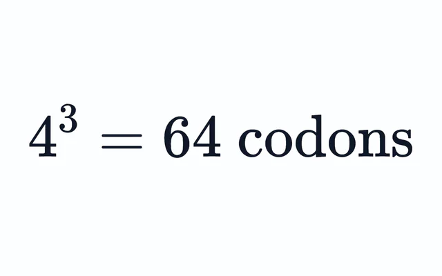
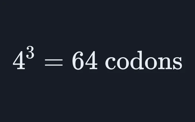
The genetic code has 61 codons for the 20 amino acids, so most have more than one. Leucine, serine and arginine have 6 each; methionine and tryptophan have 1 each. The Codons readout lists them as mRNA reads them. The DNA to Protein Translator turns a sequence into its amino acids, and the Protein Synthesis Simulator builds the chain on a ribosome, its beads in these class colours.
What this model leaves out
- Groups that affect each other. Each group is treated as ionising independently, the standard textbook treatment. In reality a charge on one group nudges the pKa of its neighbours, so the shares of the rarer protonation states are only approximate, and the measured pKas belong to the whole molecule, which is why tyrosine’s curve crosses zero at 5.63 rather than at its pI, 5.66.
- The rest of the protein. These are free amino acids. In a protein the α-amino and α-carboxyl groups are tied up in peptide bonds, except at the two ends, and a side chain’s pKa can shift by a unit or more with its surroundings.
- Other conformations. Each amino acid is one idealised conformer, from the wwPDB Chemical Component Dictionary, not the commonest arrangement of its side chain in proteins. The model places the α-amino hydrogens itself, as a regular tetrahedron, and draws every other atom where the dictionary puts it.
- One table. The pKa values are those of one table, for free amino acids in water. Other tables round them or give them for other temperatures and salt concentrations.
Common mistakes
- Averaging all three pKas. For lysine that gives
(2.18 + 8.95 + 10.53)/3 = 7.22, but its pI is 9.74, the mean of the two pKas either side of its neutral form. - Treating histidine as positive at pH 7.4. It is filed with the positive amino acids, but at pH 7.4 only about 4 percent of its side chains carry the proton, and its net charge is +0.02.
- Treating cysteine as neutral. Its thiol, pKR 8.18, is about 14 percent ionised at pH 7.4, so cysteine carries −0.14.
- Mixing up L with S. L and D name the arrangement; R and S come from ranking the groups. L-cysteine is R.
- Drawing the uncharged form. In water an amino acid near its pI is almost entirely the zwitterion, NH₃⁺ and COO⁻, not NH₂ and COOH: 99.4 percent of glycine at pH 7.4.
Where the numbers come from
The 3D coordinates are the ideal coordinates of the wwPDB Chemical Component Dictionary (Westbrook et al. (2015), Bioinformatics 31:1274), made available under the CC0 1.0 Universal Public Domain Dedication. Its backbone bond lengths agree with the values Engh and Huber (1991) set for protein structures to within 0.03 Å. The pKa values and pIs are from Nelson and Cox, Lehninger Principles of Biochemistry, 7th edition, Table 3-1, hydropathy from Kyte and Doolittle (1982), the space-filling radii from Bondi (1964), and the molar masses from the IUPAC standard atomic weights.
Common questions
What is the charge of each amino acid at pH 7.4?
At pH 7.4, the pH of blood plasma, aspartic acid and glutamic acid carry about −1 (−1.01 and −1.00) and lysine and arginine about +1 (+0.97 and +0.98). Histidine carries only +0.02: its side chain’s pKa is 6.00, so only about 4 percent of its side chains hold a proton. Cysteine carries −0.14, because its thiol, pKa 8.18, is about 14 percent ionised. The other 14 are within 0.04 of zero. The figures come from the pKa values in Lehninger’s Table 3-1, with each group treated on its own.
How do you calculate the isoelectric point of an amino acid?
Average the two pKas either side of the neutral form. For glycine that is (2.34 + 9.60)/2 = 5.97. Aspartic acid’s neutral form lies between its two carboxyl pKas, so its pI is (1.88 + 3.65)/2 ≈ 2.77, and lysine’s lies between its two amino pKas, so its pI is (8.95 + 10.53)/2 = 9.74. Averaging all three pKas gives the wrong answer. The explorer’s net charge curve, which treats each group on its own, crosses zero within 0.01 of the rule for every amino acid but tyrosine: 5.63 against the table’s 5.66. There the table is right: tyrosine’s α-amino and phenol pKas, 9.11 and 10.07, are close enough that treating the groups separately counts about a tenth too many negative molecules near its pI.
How are the 20 amino acids classified by side chain?
Lehninger’s textbook uses five classes. Nonpolar, aliphatic: glycine, alanine, proline, valine, leucine, isoleucine and methionine. Aromatic: phenylalanine, tyrosine and tryptophan. Polar, uncharged: serine, threonine, cysteine, asparagine and glutamine. Positively charged: lysine, arginine and histidine. Negatively charged: aspartate and glutamate. Some books move glycine, cysteine, histidine or tyrosine to another class, and histidine in particular carries only +0.02 at pH 7.4.
Why are the amino acids in proteins L, and why is L-cysteine R?
Every chiral amino acid a ribosome builds into a protein is the L form, the arrangement that matches L-glyceraldehyde; why life settled on L rather than D is still an open question. L and R or S are different labels. By the Cahn-Ingold-Prelog rules the α-carbon of 18 of the 19 chiral amino acids is S, because NH₂ outranks COOH, which outranks the side chain. In cysteine the sulfur in CH₂SH outranks the oxygens of COOH, so the side chain ranks second and L-cysteine is R, with the same shape.
What is a zwitterion?
A molecule carrying a positive and a negative charge that cancel. An amino acid whose side chain does not ionise is mostly zwitterion between its two pKas, and almost all zwitterion well inside them, with NH₃⁺ and COO⁻: at pH 7.4 about 99.4 percent of glycine is in that form. Open an amino acid with hydrogens on and the α-amino group shows its third hydrogen, while the carboxyl hydrogen has faded away. The uncharged form, with NH₂ and COOH, is only a tiny fraction at any pH.
Why do pKa values differ between textbooks?
They were measured at different temperatures and salt concentrations, and many tables round them: histidine’s side chain is often given as 6.0, and some tables give cysteine’s as 8.3. Inside a protein a side chain’s pKa can shift by a pH unit or more with its surroundings. The explorer uses one table, Lehninger’s Table 3-1, and names it in its readouts.
Which amino acids are the most hydrophobic?
On Kyte and Doolittle’s hydropathy scale, isoleucine (4.5), valine (4.2) and leucine (3.8) are the most hydrophobic, followed by phenylalanine (2.8) and cysteine (2.5). Arginine (−4.5) is the most hydrophilic, then lysine (−3.9). Hydrophobic side chains tend to end up inside a folded protein. The explorer shows each value in its Hydropathy readout.