Extensor digitorum
The extensor digitorum is a superficial muscle of the posterior forearm that divides into four tendons and extends the index, middle, ring and little fingers. (StatPearls, Wrist Extensor Muscles; Gray’s Anatomy (1918), Dorsal Antibrachial Muscles)
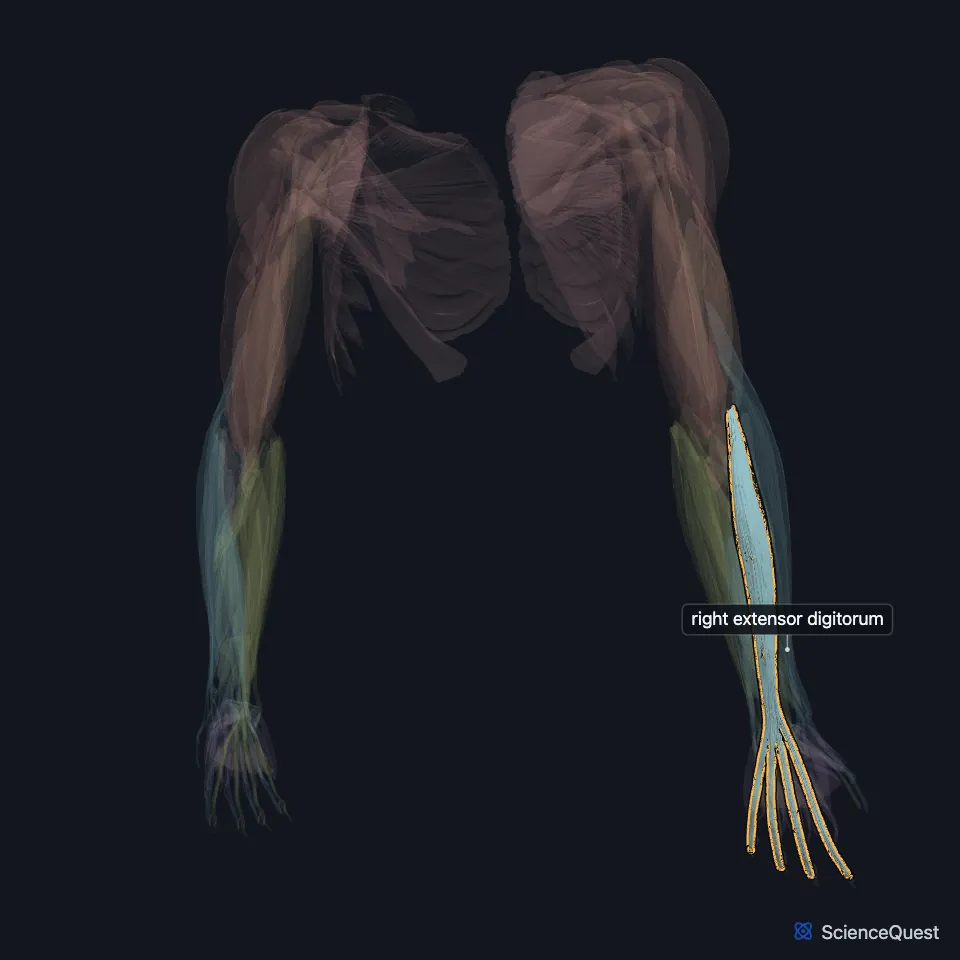
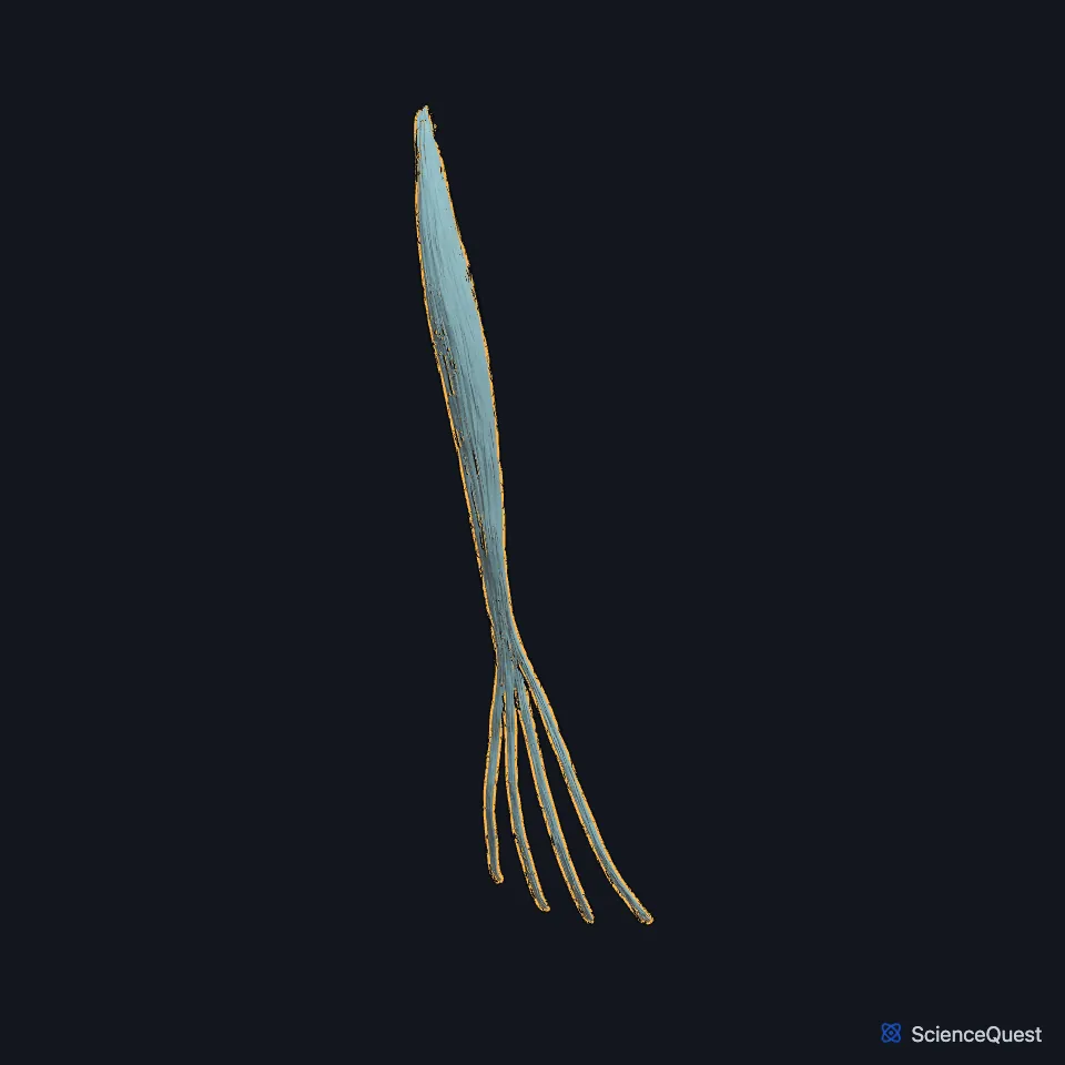
Open it in the Upper Limb Muscles Explorer See it alone in 3D
Where is the extensor digitorum?
It lies in the superficial layer of the back of the forearm. The extensor digiti minimi lies on its medial side and is usually joined to it. (StatPearls, Wrist Extensor Muscles; Gray’s Anatomy (1918), Dorsal Antibrachial Muscles)
Its four tendons pass under the extensor retinaculum in the fourth dorsal compartment, together with the tendon of the extensor indicis. (StatPearls, Wrist Extensor Muscles; Gray’s Anatomy (1918), Dorsal Antibrachial Muscles)
How do the extensor digitorum tendons attach?
It arises from the lateral epicondyle of the humerus by the common extensor tendon. Over the finger, each tendon splits into a middle slip to the base of the middle phalanx and two side slips that join and insert on the base of the distal phalanx. (Gray’s Anatomy (1918), Dorsal Antibrachial Muscles; StatPearls, Forearm Muscles)
On the back of the hand two oblique bands link the tendons to the middle, ring and little fingers. These connections limit independent extension, and they can keep a knuckle extending even after one tendon is cut. (Gray’s Anatomy (1918), Dorsal Antibrachial Muscles; StatPearls, Hand Extensor Tendon Lacerations)
What does the extensor digitorum do?
It extends the four fingers, mainly at the metacarpophalangeal joints, and helps extend the interphalangeal joints through the extensor mechanism. Gray’s 1918 adds that it spreads the fingers as it extends them, and then extends the wrist. (StatPearls, Wrist Extensor Muscles; Gray’s Anatomy (1918), Dorsal Antibrachial Muscles)
Gray’s 1918 assigns most extension of the middle and distal phalanges to the interossei and lumbricals. (Gray’s Anatomy (1918), Dorsal Antibrachial Muscles)
Which nerve supplies the extensor digitorum, and what can press on its tendons?
The posterior interosseous nerve supplies it. After crossing the supinator, the nerve divides, and its superficial group of branches supplies this muscle, the extensor digiti minimi and the extensor carpi ulnaris. (StatPearls, Hand Extensor Pollicis Longus Muscle)
Ganglion cysts, the most common soft-tissue masses of the wrist, can form on the back of the wrist and press on its tendons, causing pain. (StatPearls, Wrist Extensor Muscles)
How does the extensor digitorum vary?
The number of its tendons varies: slips to a finger may split or be absent, and Gray’s 1918 notes an occasional extra slip to the thumb. (StatPearls, Wrist Extensor Muscles; Gray’s Anatomy (1918), Dorsal Antibrachial Muscles)
| Feature | Detail and source |
|---|---|
| Origin | Lateral epicondyle of the humerus (common extensor tendon) (Gray’s Anatomy (1918), Dorsal Antibrachial Muscles; StatPearls, Wrist Extensor Muscles) |
| Insertion | Extensor expansions of the index, middle, ring and little fingers (StatPearls, Wrist Extensor Muscles) |
| Action | Extends the fingers, mainly at the metacarpophalangeal joints (StatPearls, Wrist Extensor Muscles) |
| Nerve | Posterior interosseous nerve (StatPearls, Hand Extensor Pollicis Longus Muscle) |
Touches in this model
Measured on the BodyParts3D meshes: surfaces within 2 mm of the extensor digitorum, nearest first, 12 of 31.
- Humeral head of right extensor carpi ulnaris in contact
- Right abductor pollicis longus in contact
- Right anconeus in contact
- Right extensor carpi radialis brevis in contact
- Right extensor digiti minimi in contact
- Right extensor indicis in contact
- Right extensor pollicis longus in contact
- Set of dorsal interossei of right hand in contact
- Set of lumbricals of right hand in contact
- Middle phalanx of right index finger in contact
- Middle phalanx of right little finger in contact
- Middle phalanx of right middle finger in contact
Sources
Sources read
- StatPearls, Anatomy, Shoulder and Upper Limb, Wrist Extensor Muscles. Nicole Reeves; Tyler Crow; Matthew A. Varacallo. Last update . CC BY-NC-ND 4.0. Read .
- Anatomy of the Human Body (Gray’s Anatomy), 20th US edition, 1918, Myology, The Muscles and Fasciae of the Forearm, 2. The Dorsal Antibrachial Muscles, pages 451 to 456; Internet Archive scan of the 1918 Lea and Febiger printing, terminology of 1918. Henry Gray; edited by Warren H. Lewis. Public domain. Read .
- StatPearls, Anatomy, Shoulder and Upper Limb, Forearm Muscles. Brittney Mitchell; Lacey Whited. Last update . CC BY-NC-ND 4.0. Read .
- StatPearls, Hand Extensor Tendon Lacerations. Bryan G. Beutel; Kristof S. Gutowski; Raghavendra Marappa-Ganeshan. Last update . CC BY-NC-ND 4.0. Read .
- StatPearls, Anatomy, Shoulder and Upper Limb, Hand Extensor Pollicis Longus Muscle. Irfan A. Khan; Marjorie V. Launico. Last update . CC BY-NC-ND 4.0. Read .
The values are facts and free to use; this page’s selection and presentation are © 2026 ScienceQuest.