Abductor pollicis longus
The abductor pollicis longus is a deep muscle of the posterior forearm whose tendon reaches the base of the thumb. It abducts and extends the thumb at the carpometacarpal joint. (StatPearls, Wrist Extensor Muscles)
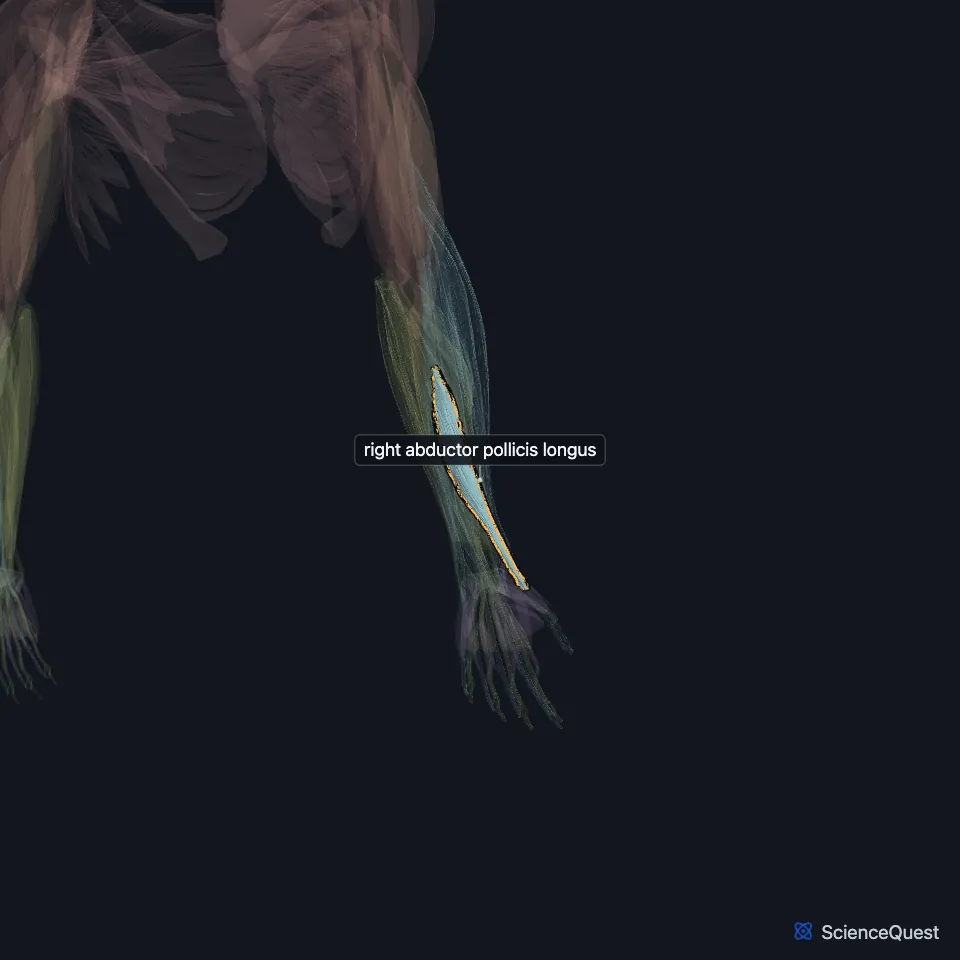
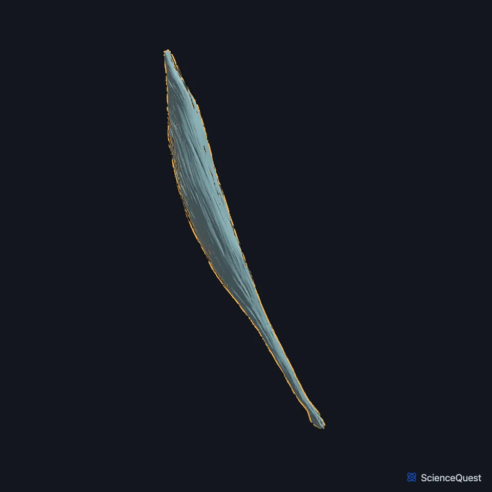
Open it in the Upper Limb Muscles Explorer See it alone in 3D
Where is the abductor pollicis longus?
It lies immediately below the supinator, which it is sometimes united with, and runs down and outward across the back of the forearm. (Gray’s Anatomy (1918), Dorsal Antibrachial Muscles)
At the wrist its tendon shares the first dorsal compartment with the extensor pollicis brevis, a fibrous tunnel about 2 cm long over the radial styloid. (StatPearls, Wrist Extensor Muscles and De Quervain Tenosynovitis)
With the extensor pollicis brevis tendon it forms the lateral border of the anatomical snuffbox. (StatPearls, Hand Anatomical Snuff Box)
Where does the abductor pollicis longus attach?
It arises from the back of the ulna, the interosseous membrane and the middle third of the back of the radius. It inserts on the radial side of the base of the first metacarpal. (Gray’s Anatomy (1918), Dorsal Antibrachial Muscles; StatPearls, Wrist Extensor Muscles)
Its tendon frequently has more than one slip. Accessory slips can insert on the trapezium, the opponens pollicis or, most commonly, the abductor pollicis brevis. (StatPearls, Wrist Extensor Muscles)
What does the abductor pollicis longus do?
It moves the thumb away from the palm, abducting and extending it at the carpometacarpal joint, and with continued action it helps extend and abduct the wrist. (Gray’s Anatomy (1918), Dorsal Antibrachial Muscles; StatPearls, Wrist Extensor Muscles)
Which nerve supplies the abductor pollicis longus, and what is de Quervain tenosynovitis?
The posterior interosseous nerve supplies it, through its deep group of branches. (StatPearls, Hand Extensor Pollicis Longus Muscle)
In de Quervain tenosynovitis the sheaths around its tendon and the extensor pollicis brevis tendon thicken in the first compartment. Thumb movement and side-to-side wrist movement make the pain worse. (StatPearls, De Quervain Tenosynovitis)
In the Finkelstein test the person holds the thumb flexed into the palm while the examiner tilts the wrist toward the ulna. Sharp pain over the first compartment is a positive result. (StatPearls, De Quervain Tenosynovitis)
How does the abductor pollicis longus vary?
A septum that splits the first compartment into separate tunnels for it and the extensor pollicis brevis is reported in more than 60% of people. If the surgeon misses a separate tunnel, de Quervain symptoms can persist after release. (StatPearls, Wrist Extensor Muscles)
Where the sources differ
Sources differ on how usual a split tendon is. The StatPearls snuffbox article gives two slips, to the base of the first metacarpal and to the trapezium, as its insertion; Gray’s 1918 says it occasionally gives off two slips near the insertion, to the greater multangular (its name for the trapezium) and to the origin of the abductor pollicis brevis. (StatPearls, Hand Anatomical Snuff Box; Gray’s Anatomy (1918), Dorsal Antibrachial Muscles)
| Feature | Detail and source |
|---|---|
| Origin | Back of the ulna and radius; interosseous membrane (StatPearls, Wrist Extensor Muscles; Gray’s Anatomy (1918), Dorsal Antibrachial Muscles) |
| Insertion | Radial side of the base of the first metacarpal (Gray’s Anatomy (1918), Dorsal Antibrachial Muscles; StatPearls, Wrist Extensor Muscles) |
| Action | Abducts and extends the thumb at the carpometacarpal joint (StatPearls, Wrist Extensor Muscles) |
| Nerve | Posterior interosseous nerve (StatPearls, Hand Extensor Pollicis Longus Muscle) |
Touches in this model
Measured on the BodyParts3D meshes: surfaces within 2 mm of the abductor pollicis longus, nearest first, 12 of 18.
- Humeral head of right extensor carpi ulnaris in contact
- Right extensor carpi radialis brevis in contact
- Right extensor carpi radialis longus in contact
- Right extensor digiti minimi in contact
- Right extensor digitorum in contact
- Right extensor pollicis brevis in contact
- Right extensor pollicis longus in contact
- Right flexor digitorum profundus in contact
- Right supinator in contact
- Ulnar head of right extensor carpi ulnaris in contact
- Interosseous membrane of right forearm in contact
- Right first metacarpal bone in contact
Sources
Sources read
- StatPearls, Anatomy, Shoulder and Upper Limb, Wrist Extensor Muscles. Nicole Reeves; Tyler Crow; Matthew A. Varacallo. Last update . CC BY-NC-ND 4.0. Read .
- Anatomy of the Human Body (Gray’s Anatomy), 20th US edition, 1918, Myology, The Muscles and Fasciae of the Forearm, 2. The Dorsal Antibrachial Muscles, pages 451 to 456; Internet Archive scan of the 1918 Lea and Febiger printing, terminology of 1918. Henry Gray; edited by Warren H. Lewis. Public domain. Read .
- StatPearls, De Quervain Tenosynovitis. Ellen Satteson; Shruti C. Tannan. Last update . CC BY-NC-ND 4.0. Read .
- StatPearls, Anatomy, Shoulder and Upper Limb: Hand Anatomical Snuff Box. Sasha Hallett Reid; Felix Jozsa; John V. Ashurst. Last update . CC BY-NC-ND 4.0. Read .
- StatPearls, Anatomy, Shoulder and Upper Limb, Hand Extensor Pollicis Longus Muscle. Irfan A. Khan; Marjorie V. Launico. Last update . CC BY-NC-ND 4.0. Read .
The values are facts and free to use; this page’s selection and presentation are © 2026 ScienceQuest.