Metacarpal bones
The five metacarpals form the skeleton of the palm, between the carpal bones and the phalanges of the fingers. They are numbered one to five from the thumb side. (StatPearls, Fifth Metacarpal Fracture; Gray’s Anatomy (1918), Metacarpus, p. 227)
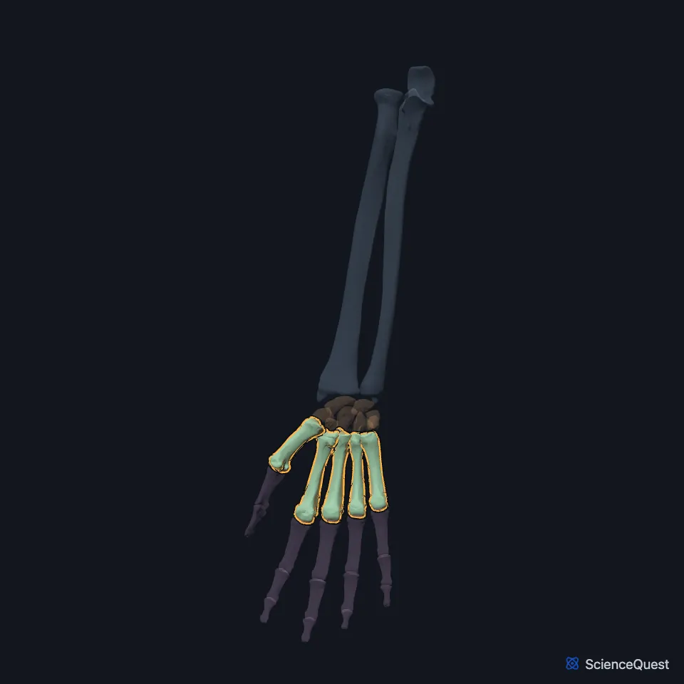
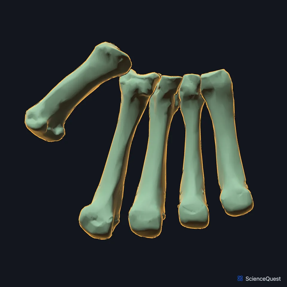
Open it in the Hand and Wrist Skeleton Explorer See it alone in 3D
Where are the metacarpals?
Each finger has one, from the first, under the thumb, to the fifth, under the little finger. Their bases meet the carpals at the carpometacarpal joints, and their heads meet the proximal phalanges at the metacarpophalangeal joints. (StatPearls, Fifth Metacarpal Fracture and Hand Bones)
Which carpal bones does each metacarpal meet?
The first meets the trapezium. The second meets the trapezium, trapezoid and capitate, the third the capitate, the fourth the capitate and hamate, and the fifth the hamate. (StatPearls, Hand Bones; Gray’s Anatomy (1918), Metacarpus, p. 227)
Tendons of the forearm end on their bases: extensor carpi radialis longus on the back of the second, flexor carpi radialis on its front, extensor carpi radialis brevis on the third, extensor carpi ulnaris on the fifth and abductor pollicis longus on the first. (Gray’s Anatomy (1918), Metacarpus, p. 227)
How do the five metacarpals differ?
Each has a base, a shaft, a neck and a head. The first is shorter and stouter than the others and angles further away from the carpus; the second is the longest, with the largest base, and the third carries a styloid process on the back of its base. (StatPearls, Fifth Metacarpal Fracture; Gray’s Anatomy (1918), Metacarpus, p. 227)
Two sesamoid bones sit at the knuckle joint of the thumb, at the far end of the first metacarpal, within the tendons of flexor pollicis brevis and adductor pollicis. (StatPearls, Hand Bones and Sesamoid Bones)
What do the metacarpals do?
The metacarpals are the bony base of the lever system the hand’s flexor and extensor tendons work on. The interosseous muscles that spread and close the fingers arise from their shafts. (StatPearls, Metacarpal Fracture and Fifth Metacarpal Fracture)
The second to fifth metacarpals are held close together, so a single broken one is often stable. (StatPearls, Fifth Metacarpal Fracture)
How do the metacarpals break?
A boxer’s fracture is a break across the neck of the fifth metacarpal, typically from punching with a clenched fist. It makes up about 10% of all hand fractures. (StatPearls, Fifth Metacarpal Fracture)
A Bennett fracture runs into the joint at the base of the first metacarpal, leaving a small fragment on the palm and little-finger side, and is often unstable. (StatPearls, Bennett Fracture)
The index and middle finger metacarpals move little where they join the trapezoid and capitate, and a fracture of one can only tolerate 10 to 15 degrees of angulation. The ring and little finger metacarpals move more on the hamate and may tolerate 30 to 45 degrees in the ring finger and up to 50 in the little finger. (StatPearls, Metacarpal Fracture)
Where the sources differ
Gray’s Anatomy (1918) puts both thumb sesamoids in the tendons of flexor pollicis brevis. StatPearls’ article on sesamoid bones places them within the tendons of flexor pollicis brevis and adductor pollicis. (Gray’s Anatomy (1918), Metacarpus, p. 227; StatPearls, Sesamoid Bones)
StatPearls’ article on fifth metacarpal fractures puts metacarpal fractures at 40% of all hand fractures. Its article on the carpal bones gives 34% of adult hand fractures. (StatPearls, Fifth Metacarpal Fracture and Hand Carpal Bones)
| Feature | Detail and source |
|---|---|
| Number | Five, numbered from the thumb side (StatPearls, Hand Bones; Gray’s Anatomy (1918), Metacarpus, p. 227) |
| Parts | Base, shaft, neck, head (StatPearls, Fifth Metacarpal Fracture) |
| Longest | Second (Gray’s Anatomy (1918), Metacarpus, p. 227) |
Touches in this model
Measured on the BodyParts3D meshes: surfaces within 2 mm of the metacarpal bones, nearest first, 12 of 37.
- Proximal phalanx of right index finger in contact
- Proximal phalanx of right little finger in contact
- Proximal phalanx of right middle finger in contact
- Proximal phalanx of right ring finger in contact
- Proximal phalanx of right thumb in contact
- Right capitate in contact
- Right hamate in contact
- Right trapezium in contact
- Right trapezoid in contact
- Deep head of right flexor pollicis brevis in contact
- Humeral head of right extensor carpi ulnaris in contact
- Humeral head of right flexor carpi ulnaris in contact
Sources
Sources read
- StatPearls, Fifth Metacarpal Fracture. Saloni Malik; Tom Herron; Muhammad Taqi; Naomi Rosenberg. Last update . CC BY-NC-ND 4.0. Read .
- Gray’s Anatomy of the Human Body, 20th US edition (1918), edited by Warren H. Lewis, Osteology, The Metacarpus (pp. 227 to 229); 1918 names: greater and lesser multangular for trapezium and trapezoid. Henry Gray; Warren H. Lewis (editor). Public domain. Read .
- StatPearls, Anatomy, Shoulder and Upper Limb, Hand Bones. Daniel G. Arias; Asa C. Black; Matthew A. Varacallo. Last update . CC BY-NC-ND 4.0. Read .
- StatPearls, Anatomy, Sesamoid Bones. Anthony Y. Yeung; Tafline C. Arbor; Dr. Rohin Garg. Last update . CC BY-NC-ND 4.0. Read .
- StatPearls, Metacarpal Fracture. Ryan L. Werntz; Matthew A. Varacallo. Last update . CC BY-NC-ND 4.0. Read .
- StatPearls, Bennett Fracture. Bryan G. Beutel; Mark Gamadia; Kevin R. Carter. Last update . CC BY-NC-ND 4.0. Read .
- StatPearls, Anatomy, Shoulder and Upper Limb, Hand Carpal Bones. Alex Tang; Matthew A. Varacallo. Last update . CC BY-NC-ND 4.0. Read .
The values are facts and free to use; this page’s selection and presentation are © 2026 ScienceQuest.