Trapezium
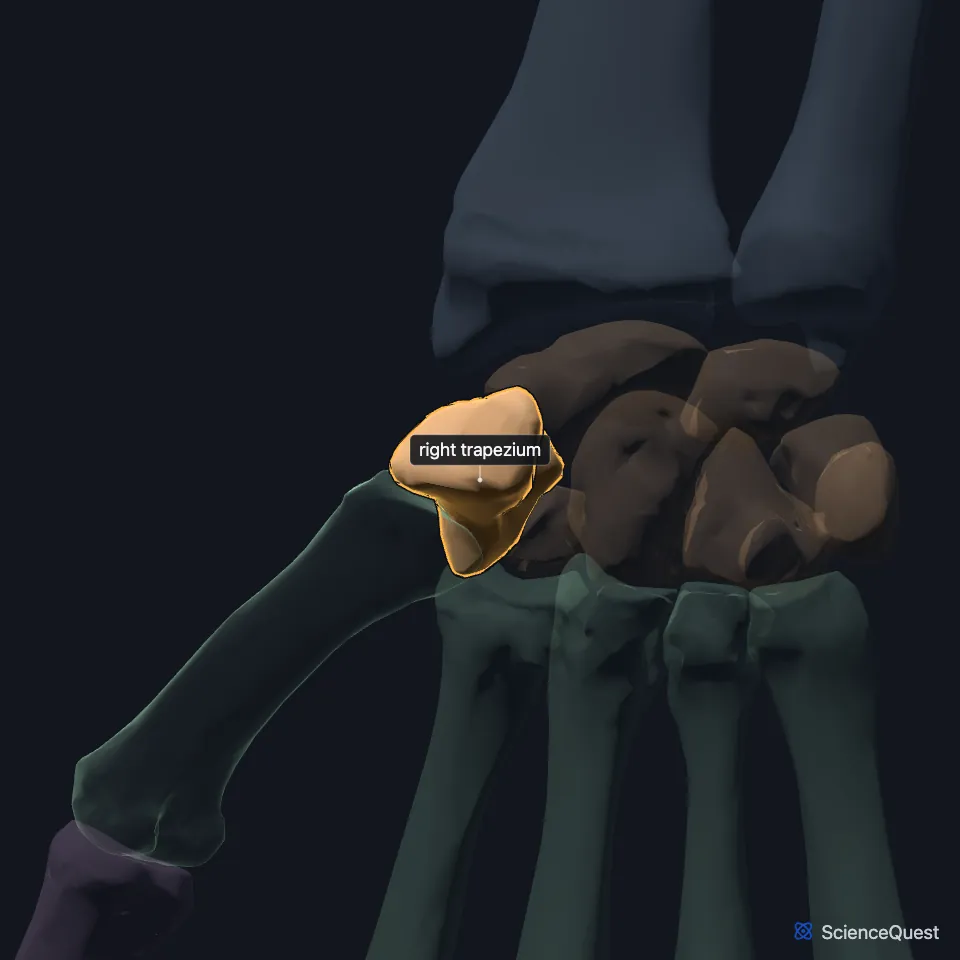
The trapezium is the carpal bone at the thumb end of the distal row. It sits between the scaphoid and the base of the first metacarpal. (StatPearls, Hand Carpal Bones; Gray’s Anatomy (1918), Greater multangular bone, p. 225)
Open it in the Hand and Wrist Skeleton Explorer See it alone in 3D
Where is the trapezium?
The distal carpal row runs trapezium, trapezoid, capitate and hamate, from the thumb side. The trapezium lies beyond the scaphoid, at the base of the thumb. (StatPearls, Hand Carpal Bones)
What is the thumb’s saddle joint?
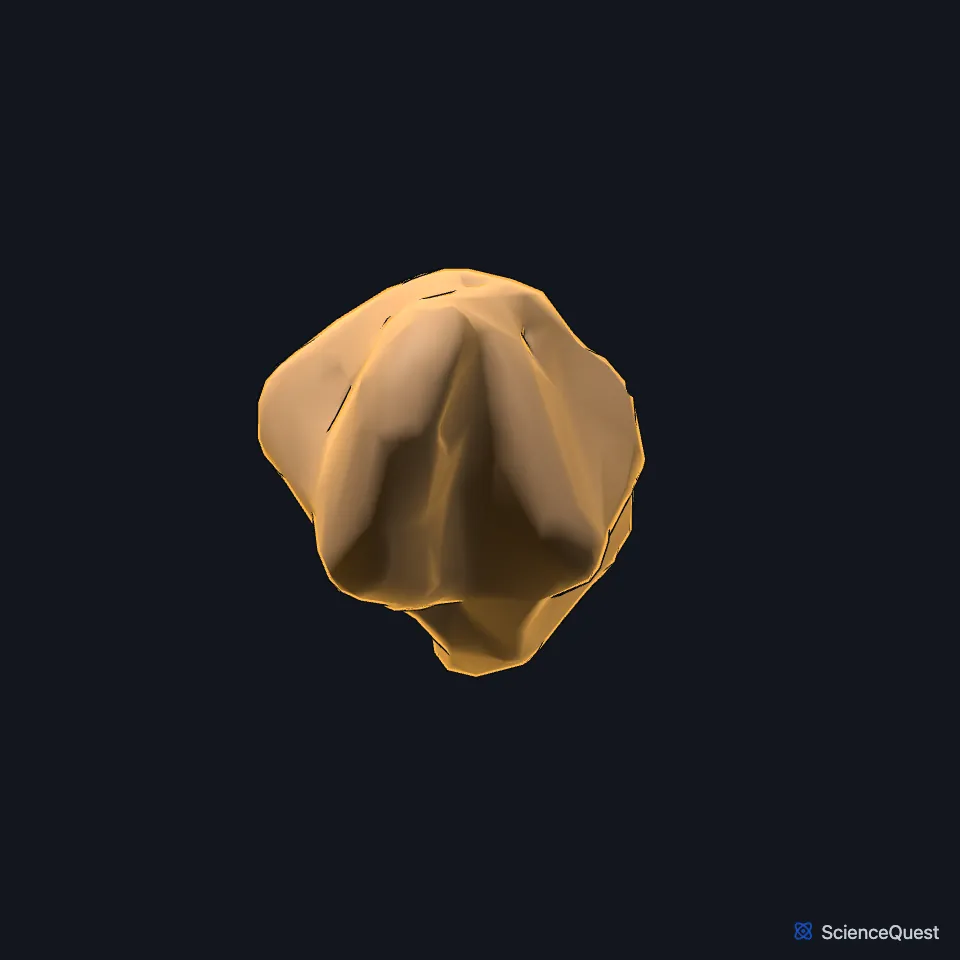
The trapezium’s lower surface is saddle-shaped, and with the base of the first metacarpal it forms the carpometacarpal joint of the thumb. That joint carries the thumb’s flexion and extension and its abduction and adduction, with little axial rotation. (StatPearls, Hand Carpal Bones and Bennett Fracture; Gray’s Anatomy (1918), Greater multangular bone, p. 225)
Its tubercle and the scaphoid’s project forward. They shape the carpal tunnel and give the thumb a base from which it opposes the other fingers. (StatPearls, Hand Carpal Bones)
Which bones and soft tissues attach to the trapezium?
It meets four bones: the scaphoid above, the first metacarpal below, and the trapezoid and second metacarpal on its inner side. Its palm surface carries a deep groove for the tendon of flexor carpi radialis. (Gray’s Anatomy (1918), Greater multangular bone, p. 225; StatPearls, Hand Bones)
All three thenar muscles take part of their origin from the trapezium. Two ligaments from the trapezium, the volar oblique and the dorsoradial, steady the base of the first metacarpal. (StatPearls, Hand Thenar Eminence and Bennett Fracture)
Which injuries involve the trapezium?
A Bennett fracture breaks the base of the first metacarpal into its joint with the trapezium, and it may come with a fracture of the trapezium itself. A fracture of the trapezium or the first metacarpal can disrupt the thenar muscles. (StatPearls, Bennett Fracture and Hand Thenar Eminence)
| Feature | Detail and source |
|---|---|
| Position | Distal carpal row, thumb end (StatPearls, Hand Carpal Bones; Gray’s Anatomy (1918), Greater multangular bone, p. 225) |
| Articulates with | Scaphoid, first and second metacarpals, trapezoid (Gray’s Anatomy (1918), Greater multangular bone, p. 225) |
| Older name | Greater multangular bone (Gray’s Anatomy, 1918) (Gray’s Anatomy (1918), Greater multangular bone, p. 225) |
Touches in this model
Measured on the BodyParts3D meshes: surfaces within 2 mm of the trapezium, nearest first, 12 of 13.
- Right first metacarpal bone in contact
- Right scaphoid in contact
- Right second metacarpal bone in contact
- Right trapezoid in contact
- Deep head of right flexor pollicis brevis in contact
- Flexor retinaculum of right wrist in contact
- Right abductor pollicis brevis in contact
- Right extensor carpi radialis longus in contact
- Right opponens pollicis in contact
- Skin in contact
- Right flexor pollicis longus 0.8 mm away
- Right extensor pollicis longus 1.4 mm away
Sources
Sources read
- StatPearls, Anatomy, Shoulder and Upper Limb, Hand Carpal Bones. Alex Tang; Matthew A. Varacallo. Last update . CC BY-NC-ND 4.0. Read .
- Gray’s Anatomy of the Human Body, 20th US edition (1918), edited by Warren H. Lewis, Osteology, The Carpus, The Greater Multangular Bone (p. 225); 1918 names: greater multangular for trapezium, navicular for scaphoid, lesser multangular for trapezoid. Henry Gray; Warren H. Lewis (editor). Public domain. Read .
- StatPearls, Bennett Fracture. Bryan G. Beutel; Mark Gamadia; Kevin R. Carter. Last update . CC BY-NC-ND 4.0. Read .
- StatPearls, Anatomy, Shoulder and Upper Limb, Hand Bones. Daniel G. Arias; Asa C. Black; Matthew A. Varacallo. Last update . CC BY-NC-ND 4.0. Read .
- StatPearls, Anatomy, Shoulder and Upper Limb, Hand Thenar Eminence. Dylan Luxenburg; Michael G. Rizzo. Last update . CC BY-NC-ND 4.0. Read .
The values are facts and free to use; this page’s selection and presentation are © 2026 ScienceQuest.