Abductor pollicis brevis
The abductor pollicis brevis is one of the three thenar muscles, the group that makes the fleshy mound at the base of the thumb on the palm side of the hand. (StatPearls, Hand Thenar Eminence)
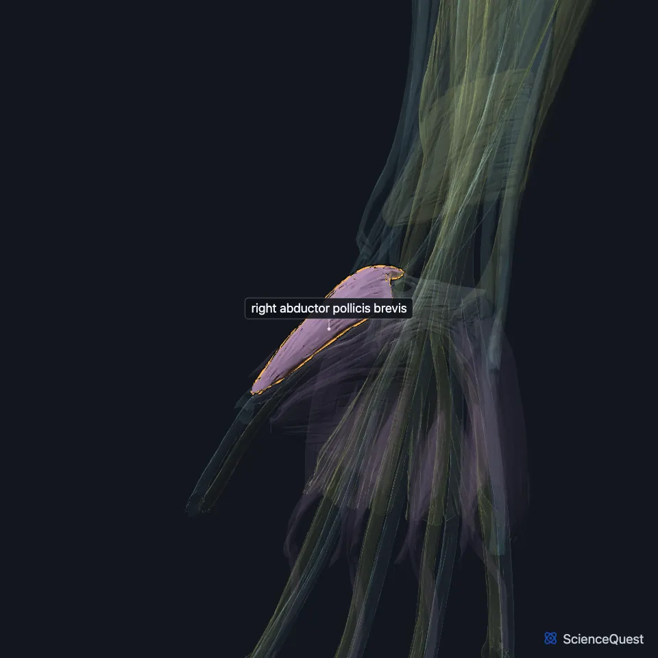
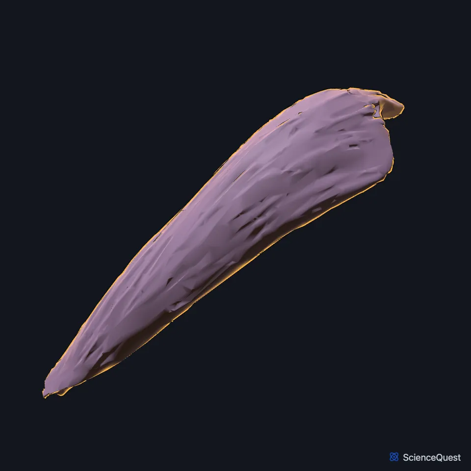
Open it in the Upper Limb Muscles Explorer See it alone in 3D
Where is the abductor pollicis brevis?
It is the most superficial thenar muscle, lying just under the skin and to the outer side of the other two. The opponens pollicis lies beneath it. (StatPearls, Hand Thenar Eminence; Gray’s Anatomy (1918), Lateral Volar Muscles)
What does the abductor pollicis brevis attach to?
It arises mainly from the flexor retinaculum, with fibers from the tubercles of the scaphoid and the trapezium. It runs down and outward to the base of the proximal phalanx of the thumb. (StatPearls, Hand Thenar Eminence)
Gray’s 1918 gives the same origin in older names: the transverse carpal ligament, the tuberosity of the navicular and the ridge of the greater multangular, often by two slips. The FMA lists navicular bone of hand as a synonym of the scaphoid, and greater multangular bone as one of the trapezium. Many experts treat the transverse carpal ligament and the flexor retinaculum as one. (Gray’s Anatomy (1918), Lateral Volar Muscles; FMA, Scaphoid and Trapezium; StatPearls, Wrist Flexor Retinaculum)
What does the abductor pollicis brevis do?
It abducts the thumb. Gray’s 1918 describes the movement as drawing the thumb forward, at right angles to the plane of the palm. (StatPearls, Hand Thenar Eminence; Gray’s Anatomy (1918), Lateral Volar Muscles)
The thumb sits rotated 90° at its carpometacarpal joint, so its movements run in other planes than the fingers’. Thumb abduction and adduction happen in the sagittal plane. (StatPearls, Wrist Flexor Retinaculum)
Which nerve and vessels supply the abductor pollicis brevis?
The recurrent branch of the median nerve supplies it. The branch leaves the median nerve after the nerve passes through the carpal tunnel, under the flexor retinaculum, and turns back to the thenar muscles. (StatPearls, Hand Thenar Eminence and Wrist Flexor Retinaculum)
Severe carpal tunnel syndrome can waste the thenar eminence. Wasting of the thenar eminence with the hypothenar eminence spared, the split hand sign, can be the first sign of amyotrophic lateral sclerosis (ALS). (StatPearls, Hand Thenar Eminence)
The superficial palmar branch of the radial artery supplies it, as it supplies the other two thenar muscles. (StatPearls, Hand Thenar Eminence)
Where the sources differ
Sources give different spinal roots for its nerve. Gray’s 1918 names the sixth and seventh cervical nerves, through the median nerve; the StatPearls thenar article gives T1 for the recurrent branch, and its opponens pollicis article gives C8 and T1. (Gray’s Anatomy (1918), Lateral Volar Muscles; StatPearls, Hand Thenar Eminence and Hand Opponens Pollicis Muscle)
| Feature | Detail and source |
|---|---|
| Origin | Flexor retinaculum; tubercles of the scaphoid and trapezium (StatPearls, Hand Thenar Eminence) |
| Insertion | Base of the proximal phalanx of the thumb (StatPearls, Hand Thenar Eminence) |
| Action | Abducts the thumb (StatPearls, Hand Thenar Eminence) |
| Nerve | Recurrent branch of the median nerve (StatPearls, Hand Thenar Eminence) |
Touches in this model
Measured on the BodyParts3D meshes: surfaces within 2 mm of the abductor pollicis brevis, nearest first.
- Flexor retinaculum of right wrist in contact
- Right opponens pollicis in contact
- Superficial head of right flexor pollicis brevis in contact
- Right first metacarpal bone in contact
- Right scaphoid in contact
- Right trapezium in contact
- Skin in contact
- Deep head of right flexor pollicis brevis 0.1 mm away
- Right flexor carpi radialis 0.5 mm away
- Right flexor pollicis longus 1.6 mm away
Sources
Sources read
- StatPearls, Anatomy, Shoulder and Upper Limb, Hand Thenar Eminence. Dylan Luxenburg; Michael G. Rizzo. Last update . CC BY-NC-ND 4.0. Read .
- Anatomy of the Human Body (Gray’s Anatomy), 20th US edition, 1918, Myology, The Muscles and Fasciae of the Hand, 1. The Lateral Volar Muscles, pages 461 to 462; Internet Archive scan of the 1918 Lea and Febiger printing, terminology of 1918. Henry Gray; edited by Warren H. Lewis. Public domain. Read .
- Foundational Model of Anatomy, FMA23709, Scaphoid (read on EBI OLS). FMA release terms, University of Washington. Read .
- Foundational Model of Anatomy, FMA23721, Trapezium (read on EBI OLS). FMA release terms, University of Washington. Read .
- StatPearls, Anatomy, Shoulder and Upper Limb, Wrist Flexor Retinaculum. Nathaniel Deak; Asa C. Black; Bruno Bordoni. Last update . CC BY-NC-ND 4.0. Read .
- StatPearls, Anatomy, Shoulder and Upper Limb, Hand Opponens Pollicis Muscle. John D. Nguyen; Hieu Duong. Last update . CC BY-NC-ND 4.0. Read .
The values are facts and free to use; this page’s selection and presentation are © 2026 ScienceQuest.