Adductor pollicis
The adductor pollicis is a triangular, two-headed intrinsic hand muscle in the deepest muscle layer of the palm, and it adducts the thumb. (StatPearls, Hand Adductor Pollicis)
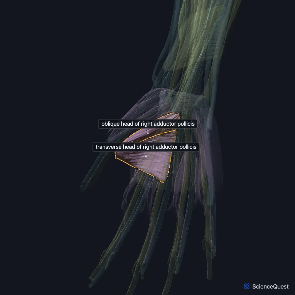
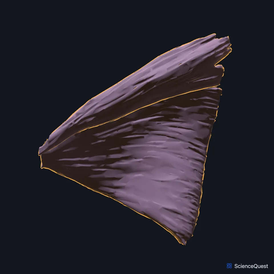
Open it in the Upper Limb Muscles Explorer See it alone in 3D
Where is the adductor pollicis?
It has a compartment of its own, the adductor compartment, apart from the thenar and hypothenar groups. The radial artery passes between its two heads to enter the deep palm and form the deep palmar arch. (StatPearls, Hand Muscles and Hand Adductor Pollicis)
What do the two heads of the adductor pollicis attach to?
The oblique head arises from the capitate and the bases of the second and third metacarpals, and the transverse head from the palm side of the third metacarpal. The heads join in a tendon, often holding a sesamoid bone, that inserts on the medial side of the thumb’s proximal phalanx and on the extensor hood. (StatPearls, Hand Adductor Pollicis)
Gray’s 1918 calls the transverse head the most deeply placed muscle of the thumb group, arising by a broad base from the lower two-thirds of the third metacarpal. (Gray’s Anatomy (1918), Lateral Volar Muscles)
What does the adductor pollicis do?
It adducts the thumb. Because the thumb lies in another plane from the palm, adduction brings it to the center of the palm, and the muscle can also bring it alongside the palm and index finger. (StatPearls, Hand Adductor Pollicis)
Pinching and gripping both need it. (StatPearls, Hand Adductor Pollicis)
Which nerve supplies the adductor pollicis, and how is it tested?
The deep branch of the ulnar nerve (C8 and T1) supplies it. The same branch supplies the hypothenar muscles, the medial two lumbricals and the interossei. (StatPearls, Hand Adductor Pollicis)
A clinician asks you to hold a sheet of paper between thumb and index finger. With a weak adductor, the thumb’s end joint bends instead, pulled by the flexor pollicis longus, which is the Froment sign of ulnar nerve weakness. (StatPearls, Hand Adductor Pollicis and Wrist Flexor Retinaculum)
Anesthesiologists stimulate the ulnar nerve and record the movements of the adductor pollicis to monitor neuromuscular blockade. (StatPearls, Hand Adductor Pollicis)
Where the sources differ
Sources differ on whether it belongs to the thenar group. The StatPearls thenar article counts three thenar muscles and notes that some include the adductor pollicis, its opponens pollicis article counts four with the adductor, and Gray’s 1918 lists it among the thumb muscles of its lateral volar group. (StatPearls, Hand Thenar Eminence and Hand Opponens Pollicis Muscle; Gray’s Anatomy (1918), Lateral Volar Muscles)
| Feature | Detail and source |
|---|---|
| Origin | Oblique head: capitate and bases of the second and third metacarpals; transverse head: palm side of the third metacarpal (StatPearls, Hand Adductor Pollicis) |
| Insertion | Medial (ulnar) side of the base of the thumb’s proximal phalanx; extensor hood (StatPearls, Hand Adductor Pollicis; Gray’s Anatomy (1918), Lateral Volar Muscles) |
| Action | Adducts the thumb (StatPearls, Hand Adductor Pollicis) |
| Nerve | Deep branch of the ulnar nerve (C8, T1) (StatPearls, Hand Adductor Pollicis) |
Touches in this model
Measured on the BodyParts3D meshes: surfaces within 2 mm of the adductor pollicis, nearest first, 12 of 16.
- Deep head of right flexor pollicis brevis in contact
- Right flexor carpi radialis in contact
- Right flexor digitorum profundus in contact
- Set of lumbricals of right hand in contact
- Set of palmar interossei of right hand in contact
- Proximal phalanx of right thumb in contact
- Right capitate in contact
- Right first metacarpal bone in contact
- Right second metacarpal bone in contact
- Right third metacarpal bone in contact
- Right trapezoid in contact
- Skin in contact
Sources
Sources read
- StatPearls, Anatomy, Shoulder and Upper Limb, Hand Adductor Pollicis. Jonathan R. Acosta; Steven B. Graefe; Matthew A. Varacallo. Last update . CC BY-NC-ND 4.0. Read .
- StatPearls, Anatomy, Shoulder and Upper Limb, Hand Muscles. Ebubechi Okwumabua; Margaret A. Sinkler; Bruno Bordoni. Last update . CC BY-NC-ND 4.0. Read .
- Anatomy of the Human Body (Gray’s Anatomy), 20th US edition, 1918, Myology, The Muscles and Fasciae of the Hand, 1. The Lateral Volar Muscles, pages 461 to 462; Internet Archive scan of the 1918 Lea and Febiger printing, terminology of 1918. Henry Gray; edited by Warren H. Lewis. Public domain. Read .
- StatPearls, Anatomy, Shoulder and Upper Limb, Wrist Flexor Retinaculum. Nathaniel Deak; Asa C. Black; Bruno Bordoni. Last update . CC BY-NC-ND 4.0. Read .
- StatPearls, Anatomy, Shoulder and Upper Limb, Hand Thenar Eminence. Dylan Luxenburg; Michael G. Rizzo. Last update . CC BY-NC-ND 4.0. Read .
- StatPearls, Anatomy, Shoulder and Upper Limb, Hand Opponens Pollicis Muscle. John D. Nguyen; Hieu Duong. Last update . CC BY-NC-ND 4.0. Read .
The values are facts and free to use; this page’s selection and presentation are © 2026 ScienceQuest.