Trapezoid
The trapezoid is a small, wedge-shaped carpal bone in the distal row, between the trapezium and the capitate. Gray’s Anatomy (1918) calls it the lesser multangular. (StatPearls, Hand Carpal Bones; Gray’s Anatomy (1918), Lesser multangular bone, p. 225)
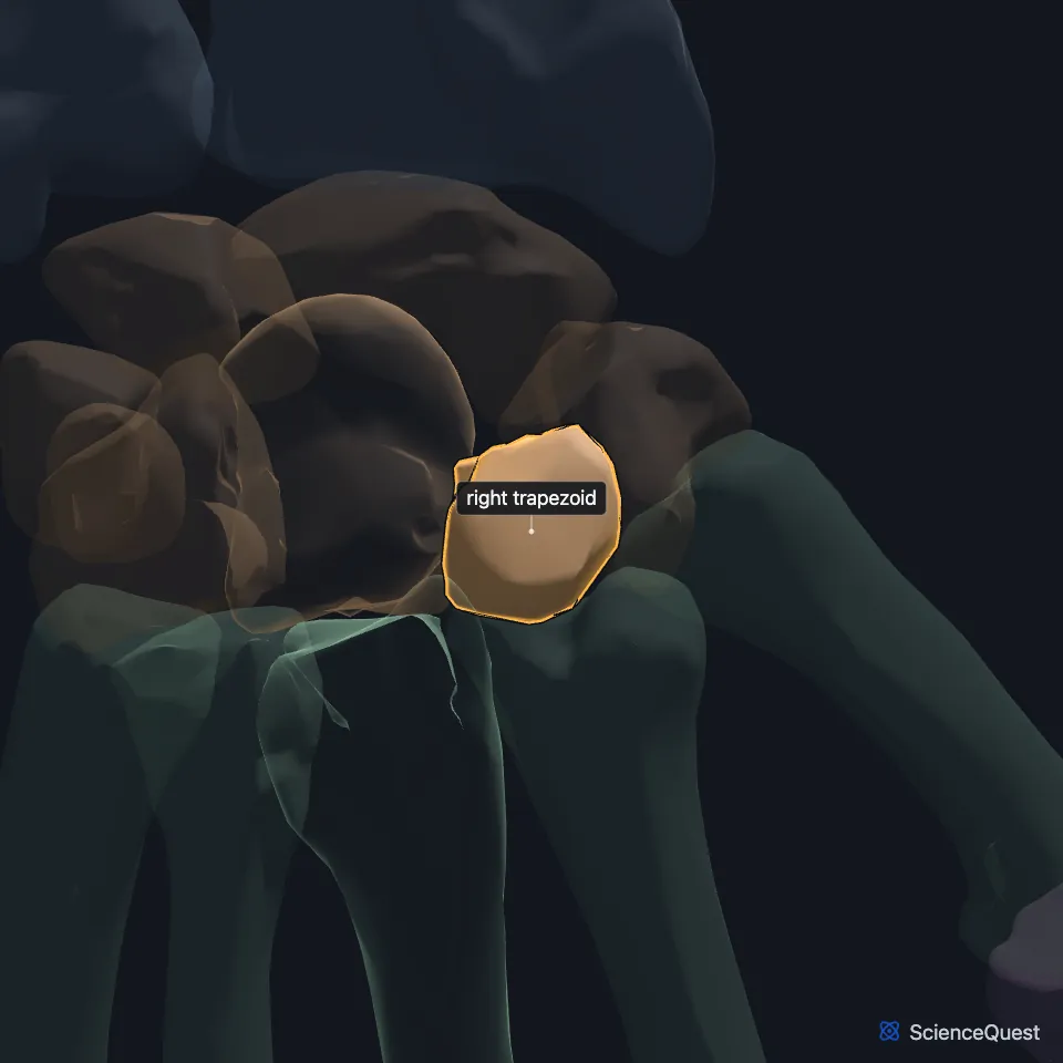
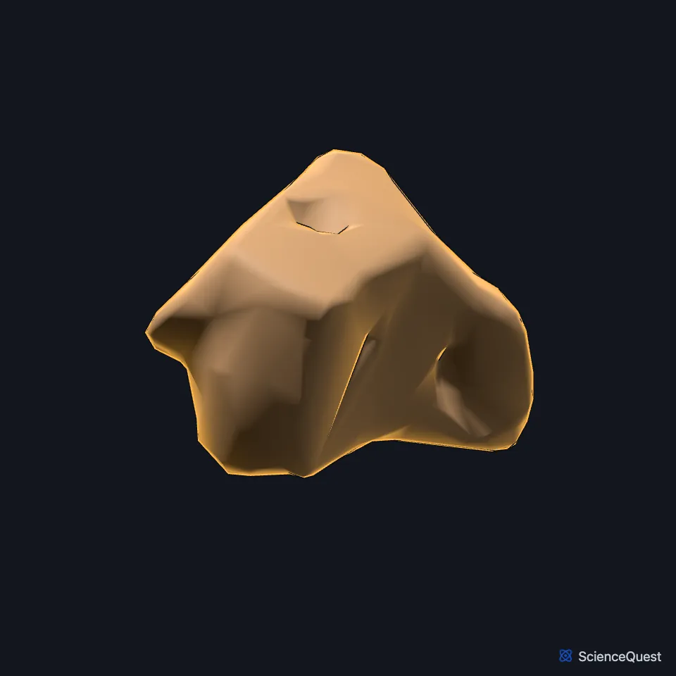
Open it in the Hand and Wrist Skeleton Explorer See it alone in 3D
Where is the trapezoid?
It is the second bone of the distal row counted from the thumb side, after the trapezium and before the capitate and hamate. The broad end of its wedge faces the back of the hand. (StatPearls, Hand Carpal Bones; Gray’s Anatomy (1918), Lesser multangular bone, p. 225)
Which bones does the trapezoid meet?
Four, through four facets that touch each other along sharp edges: the scaphoid above, the second metacarpal below, the trapezium on its thumb side and the capitate on its other side. (Gray’s Anatomy (1918), Lesser multangular bone, p. 225)
The base of the second metacarpal fits against three carpals: the trapezium, the trapezoid and the capitate. (StatPearls, Hand Bones)
What does the trapezoid do?
The distal row, the trapezoid with the trapezium, capitate and hamate, is the more rigid of the two rows and bridges the proximal row to the bases of the metacarpals. The proximal row makes most of the wrist’s movement. (StatPearls, Perilunate Dislocation and Kienbock Disease)
Because the index and middle finger metacarpals are bound to the trapezoid and capitate, they move less than the ring and little finger metacarpals. After a fracture, those two metacarpals tolerate only 10 to 15 degrees of angulation. (StatPearls, Metacarpal Fracture)
How does blood reach the trapezoid?
The trapezoid, like the hamate, has two areas of blood supply with no vessels joining them inside the bone. StatPearls counts it among the carpals at lower risk of avascular necrosis after a fracture than the scaphoid and capitate. (StatPearls, Hand Carpal Bones)
Where the sources differ
StatPearls calls the trapezoid the smallest carpal bone. Gray’s Anatomy (1918) calls it the smallest bone of the distal row, and singles out the pisiform of the proximal row by its small size. (StatPearls, Hand Carpal Bones; Gray’s Anatomy (1918), Lesser multangular bone, p. 225 and Pisiform bone, p. 225)
| Feature | Detail and source |
|---|---|
| Position | Distal carpal row, between trapezium and capitate (StatPearls, Hand Carpal Bones) |
| Articulates with | Scaphoid, second metacarpal, trapezium, capitate (Gray’s Anatomy (1918), Lesser multangular bone, p. 225) |
| Older name | Lesser multangular bone (Gray’s Anatomy, 1918) (Gray’s Anatomy (1918), Lesser multangular bone, p. 225) |
Touches in this model
Measured on the BodyParts3D meshes: surfaces within 2 mm of the trapezoid, nearest first.
- Right capitate in contact
- Right third metacarpal bone in contact
- Right trapezium in contact
- Deep head of right flexor pollicis brevis in contact
- Oblique head of right adductor pollicis in contact
- Right extensor carpi radialis brevis in contact
- Right extensor indicis in contact
- Skin in contact
- Right extensor carpi radialis longus 0.6 mm away
- Right second metacarpal bone 0.7 mm away
- Right scaphoid 1.4 mm away
- Right extensor digitorum 1.7 mm away
Sources
Sources read
- StatPearls, Anatomy, Shoulder and Upper Limb, Hand Carpal Bones. Alex Tang; Matthew A. Varacallo. Last update . CC BY-NC-ND 4.0. Read .
- Gray’s Anatomy of the Human Body, 20th US edition (1918), edited by Warren H. Lewis, Osteology, The Carpus, The Lesser Multangular Bone (pp. 225 to 226); 1918 names: lesser multangular for trapezoid, greater multangular for trapezium, navicular for scaphoid. Henry Gray; Warren H. Lewis (editor). Public domain. Read .
- StatPearls, Anatomy, Shoulder and Upper Limb, Hand Bones. Daniel G. Arias; Asa C. Black; Matthew A. Varacallo. Last update . CC BY-NC-ND 4.0. Read .
- StatPearls, Perilunate Dislocation. Nicholas Frane; William Goldenberg. Last update . CC BY-NC-ND 4.0. Read .
- StatPearls, Kienbock Disease. Layla A. Nasr; Jennifer Koay. Last update . CC BY-NC-ND 4.0. Read .
- StatPearls, Metacarpal Fracture. Ryan L. Werntz; Matthew A. Varacallo. Last update . CC BY-NC-ND 4.0. Read .
- Gray’s Anatomy of the Human Body, 20th US edition (1918), edited by Warren H. Lewis, Osteology, The Carpus, The Pisiform Bone (p. 225); 1918 names: triangular for triquetral, Abductor digiti quinti for abductor digiti minimi. Henry Gray; Warren H. Lewis (editor). Public domain. Read .
The values are facts and free to use; this page’s selection and presentation are © 2026 ScienceQuest.