Flexor carpi radialis
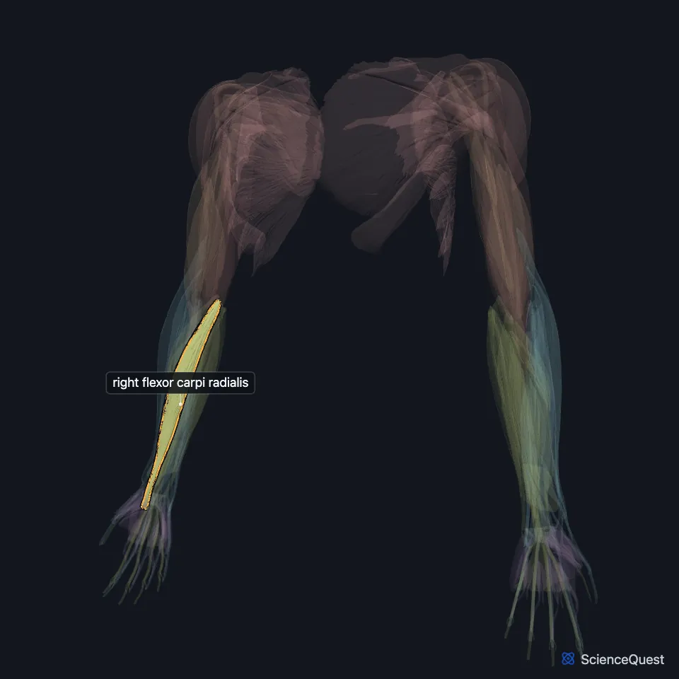
The flexor carpi radialis is one of the superficial flexor muscles on the front of the forearm. It flexes the wrist and abducts it, tilting the hand toward the thumb side. (Gray’s Anatomy (1918), Volar Antibrachial Muscles; StatPearls, Forearm Compartments)
Open it in the Upper Limb Muscles Explorer See it alone in 3D
Where is the flexor carpi radialis?
It lies on the medial side of the pronator teres, with the palmaris longus on its other side and the flexor digitorum superficialis beneath it. (Gray’s Anatomy (1918), Volar Antibrachial Muscles)
In the lower forearm the radial artery runs between its tendon and the brachioradialis, on the lateral side of the muscle. (Gray’s Anatomy (1918), Volar Antibrachial Muscles; StatPearls, Forearm Compartments)
Where does the flexor carpi radialis attach?
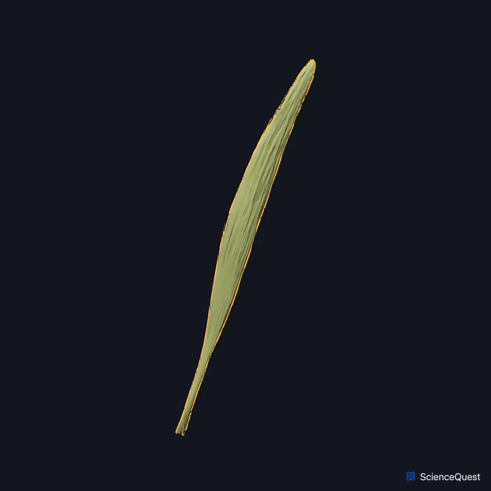
It arises from the medial epicondyle of the humerus through the common flexor tendon, and from the forearm fascia. The muscle starts slender, and its tendon makes up a little more than the lower half of its length. (Gray’s Anatomy (1918), Volar Antibrachial Muscles; StatPearls, Forearm Muscles)
Its tendon passes through a canal in the lateral part of the flexor retinaculum (the transverse carpal ligament in Gray’s 1918) and inserts on the base of the second metacarpal, sending a slip to the base of the third. (Gray’s Anatomy (1918), Volar Antibrachial Muscles; StatPearls, Hand Flexor Pollicis Longus Muscle and Forearm Compartments)
What does the flexor carpi radialis do?
It flexes and abducts the wrist. Gray’s 1918 adds that it helps pronate the hand and bend the elbow. (Gray’s Anatomy (1918), Volar Antibrachial Muscles; StatPearls, Forearm Compartments)
Golfer’s elbow (medial epicondylitis) is a tendinopathy of the common flexor origin. The examiner finds the tender point 5 to 10 mm below and in front of the medial epicondyle, often over the pronator teres and this muscle. (StatPearls, Medial Epicondylitis)
Which nerve supplies the flexor carpi radialis, and which muscles share its origin?
The median nerve supplies it at the elbow. Gray’s 1918 traces that supply mainly to the sixth cervical nerve. (StatPearls, Median Nerve; Gray’s Anatomy (1918), Volar Antibrachial Muscles)
It shares the common flexor origin on the medial epicondyle with the pronator teres, palmaris longus, flexor digitorum superficialis and the humeral head of the flexor carpi ulnaris. (StatPearls, Medial Epicondylitis)
Does the flexor carpi radialis vary?
Gray’s 1918 records insertions that vary, including the trapezium and the fourth metacarpal, and notes that the muscle may be absent. (Gray’s Anatomy (1918), Volar Antibrachial Muscles)
| Feature | Detail and source |
|---|---|
| Origin | Medial epicondyle of the humerus (common flexor tendon); forearm fascia (Gray’s Anatomy (1918), Volar Antibrachial Muscles; StatPearls, Forearm Compartments) |
| Insertion | Base of the second metacarpal, with a slip to the base of the third (Gray’s Anatomy (1918), Volar Antibrachial Muscles; StatPearls, Forearm Compartments) |
| Action | Flexes and abducts the wrist (Gray’s Anatomy (1918), Volar Antibrachial Muscles; StatPearls, Forearm Compartments) |
| Nerve | Median nerve (StatPearls, Median Nerve) |
Touches in this model
Measured on the BodyParts3D meshes: surfaces within 2 mm of the flexor carpi radialis, nearest first, 12 of 15.
- Deep head of right flexor pollicis brevis in contact
- Flexor retinaculum of right wrist in contact
- Humeral head of right pronator teres in contact
- Humeroulnar head of right flexor digitorum superficialis in contact
- Oblique head of right adductor pollicis in contact
- Radial head of right flexor digitorum superficialis in contact
- Right palmaris longus in contact
- Right humerus in contact
- Right second metacarpal bone in contact
- Skin in contact
- Right flexor pollicis longus 0.1 mm away
- Right opponens pollicis 0.4 mm away
Sources
Sources read
- Anatomy of the Human Body (Gray’s Anatomy), 20th US edition, 1918, Myology, The Muscles and Fasciae of the Forearm, 1. The Volar Antibrachial Muscles, pages 445 to 451; Internet Archive scan of the 1918 Lea and Febiger printing, terminology of 1918. Henry Gray; edited by Warren H. Lewis. Public domain. Read .
- StatPearls, Anatomy, Shoulder and Upper Limb, Forearm Compartments. Zayed L. Fazal; Ayesha M. Hafeez; Margaret A. Sinkler; Abdul Arain. Last update . CC BY-NC-ND 4.0. Read .
- StatPearls, Anatomy, Shoulder and Upper Limb, Forearm Muscles. Brittney Mitchell; Lacey Whited. Last update . CC BY-NC-ND 4.0. Read .
- StatPearls, Anatomy, Shoulder and Upper Limb, Hand Flexor Pollicis Longus Muscle. Dillon C. Benson; Kathleen H. Miao; Matthew A. Varacallo. Last update . CC BY-NC-ND 4.0. Read .
- StatPearls, Medial Epicondylitis (Golfer’s Elbow). Daniel Li; Ahmad Hammad; Kimberly Kaiser. Last update . CC BY-NC-ND 4.0. Read .
- StatPearls, Anatomy, Shoulder and Upper Limb, Median Nerve. Kelly A. Murphy; Nicholas Ryan; Adegbenro O. Fakoya. Last update . CC BY-NC-ND 4.0. Read .
The values are facts and free to use; this page’s selection and presentation are © 2026 ScienceQuest.