Flexor carpi ulnaris
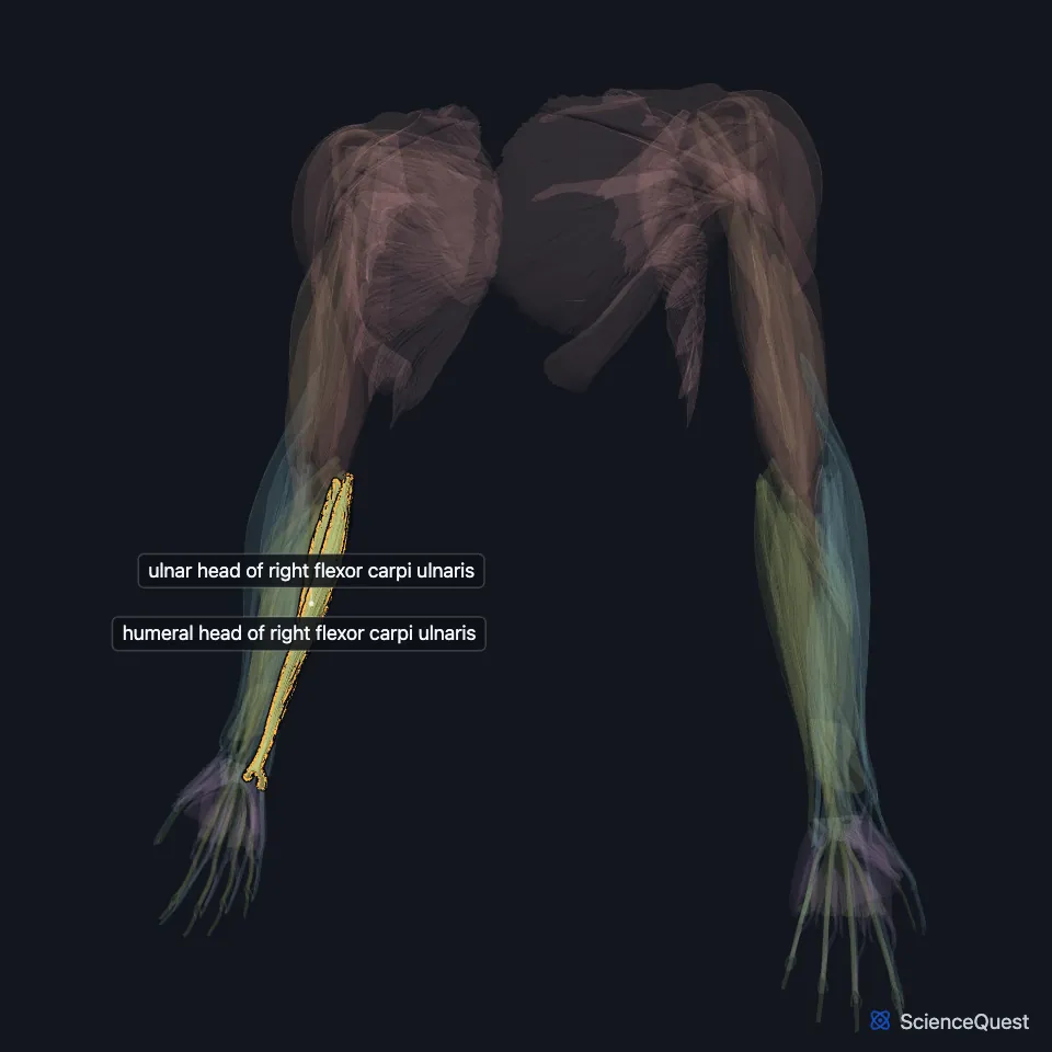
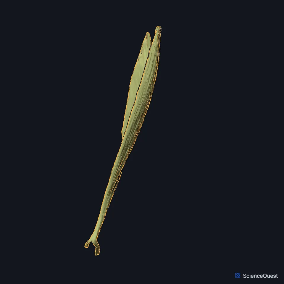
The flexor carpi ulnaris is the most medial of the superficial flexor muscles of the forearm. It is the most powerful wrist flexor, and the only muscle on the front of the forearm that the ulnar nerve supplies entirely. (StatPearls, Forearm Flexor Carpi Ulnaris Muscle)
Open it in the Upper Limb Muscles Explorer See it alone in 3D
Where is the flexor carpi ulnaris?
It runs along the ulnar side of the forearm. In the lower two-thirds of the forearm the ulnar nerve and ulnar vessels lie on the lateral side of its tendon, and surgeons use the tendon to find the nerve and artery. (Gray’s Anatomy (1918), Volar Antibrachial Muscles; StatPearls, Forearm Flexor Carpi Ulnaris Muscle)
What are the two heads of the flexor carpi ulnaris, and where does it insert?
A tendinous arch joins its two heads. The humeral head arises from the medial epicondyle through the common flexor tendon, and the ulnar head arises by an aponeurosis from the olecranon and the posterior border of the ulna. (StatPearls, Forearm Flexor Carpi Ulnaris Muscle)
Its ulnar head shares an aponeurosis on the posterior border of the ulna with the extensor carpi ulnaris and the flexor digitorum profundus. (Gray’s Anatomy (1918), Volar Antibrachial Muscles)
Its tendon inserts on the pisiform, and the pisohamate and pisometacarpal ligaments carry it on to the hook of the hamate and the fifth metacarpal. The pisiform is a sesamoid bone within the tendon. (StatPearls, Forearm Flexor Carpi Ulnaris Muscle; Gray’s Anatomy (1918), Volar Antibrachial Muscles)
What does the flexor carpi ulnaris do?
It flexes the wrist and adducts it, bending the hand toward the little finger side, and it assists flexion at the elbow. (StatPearls, Forearm Flexor Carpi Ulnaris Muscle; Gray’s Anatomy (1918), Volar Antibrachial Muscles)
How does the ulnar nerve pass the flexor carpi ulnaris?
The ulnar nerve enters the forearm between the two heads, under the arch that joins them, and gives the muscle its nerve supply. (Gray’s Anatomy (1918), Volar Antibrachial Muscles; StatPearls, Forearm Flexor Carpi Ulnaris Muscle)
The space between the two heads is the cubital tunnel. Compression of the ulnar nerve there, cubital tunnel syndrome, is the second most common compression neuropathy of the upper limb and causes tingling in the little finger and the ulnar half of the ring finger. (StatPearls, Forearm Flexor Carpi Ulnaris Muscle)
Where the sources differ
Sources give different lengths for the ulnar head’s origin. StatPearls gives the upper three-fourths of the ulna’s posterior (subcutaneous) border; Gray’s 1918 gives the upper two-thirds. (StatPearls, Forearm Flexor Carpi Ulnaris Muscle; Gray’s Anatomy (1918), Volar Antibrachial Muscles)
They also differ on its nerve roots. StatPearls gives C7 and C8; Gray’s 1918 gives the eighth cervical and first thoracic nerves. (StatPearls, Forearm Flexor Carpi Ulnaris Muscle; Gray’s Anatomy (1918), Volar Antibrachial Muscles)
| Feature | Detail and source |
|---|---|
| Origin | Humeral head: medial epicondyle (common flexor tendon). Ulnar head: olecranon and posterior border of the ulna (StatPearls, Forearm Flexor Carpi Ulnaris Muscle) |
| Insertion | Pisiform; hook of the hamate and fifth metacarpal through the pisohamate and pisometacarpal ligaments (StatPearls, Forearm Flexor Carpi Ulnaris Muscle) |
| Action | Flexes and adducts the wrist; assists elbow flexion (StatPearls, Forearm Flexor Carpi Ulnaris Muscle) |
| Nerve | Ulnar nerve (StatPearls, Forearm Flexor Carpi Ulnaris Muscle) |
Touches in this model
Measured on the BodyParts3D meshes: surfaces within 2 mm of the flexor carpi ulnaris, nearest first, 12 of 20.
- Abductor digiti minimi of right hand in contact
- Flexor digiti minimi brevis of right hand in contact
- Flexor retinaculum of right wrist in contact
- Humeral head of right extensor carpi ulnaris in contact
- Humeroulnar head of right flexor digitorum superficialis in contact
- Long head of right triceps brachii in contact
- Medial head of right triceps brachii in contact
- Right anconeus in contact
- Right palmaris longus in contact
- Right pronator quadratus in contact
- Ulnar head of right extensor carpi ulnaris in contact
- Right fifth metacarpal bone in contact
Sources
Sources read
- StatPearls, Anatomy, Shoulder and Upper Limb, Forearm Flexor Carpi Ulnaris Muscle. Brandon E. Lung; Ryan M. Siwiec. Last update . CC BY-NC-ND 4.0. Read .
- Anatomy of the Human Body (Gray’s Anatomy), 20th US edition, 1918, Myology, The Muscles and Fasciae of the Forearm, 1. The Volar Antibrachial Muscles, pages 445 to 451; Internet Archive scan of the 1918 Lea and Febiger printing, terminology of 1918. Henry Gray; edited by Warren H. Lewis. Public domain. Read .
The values are facts and free to use; this page’s selection and presentation are © 2026 ScienceQuest.