Flexor digitorum superficialis
The flexor digitorum superficialis is the largest extrinsic flexor of the forearm. It forms the intermediate layer of the front of the forearm and sends a tendon to each of the four fingers. (StatPearls, Hand Flexor Digitorum Superficialis Muscle)
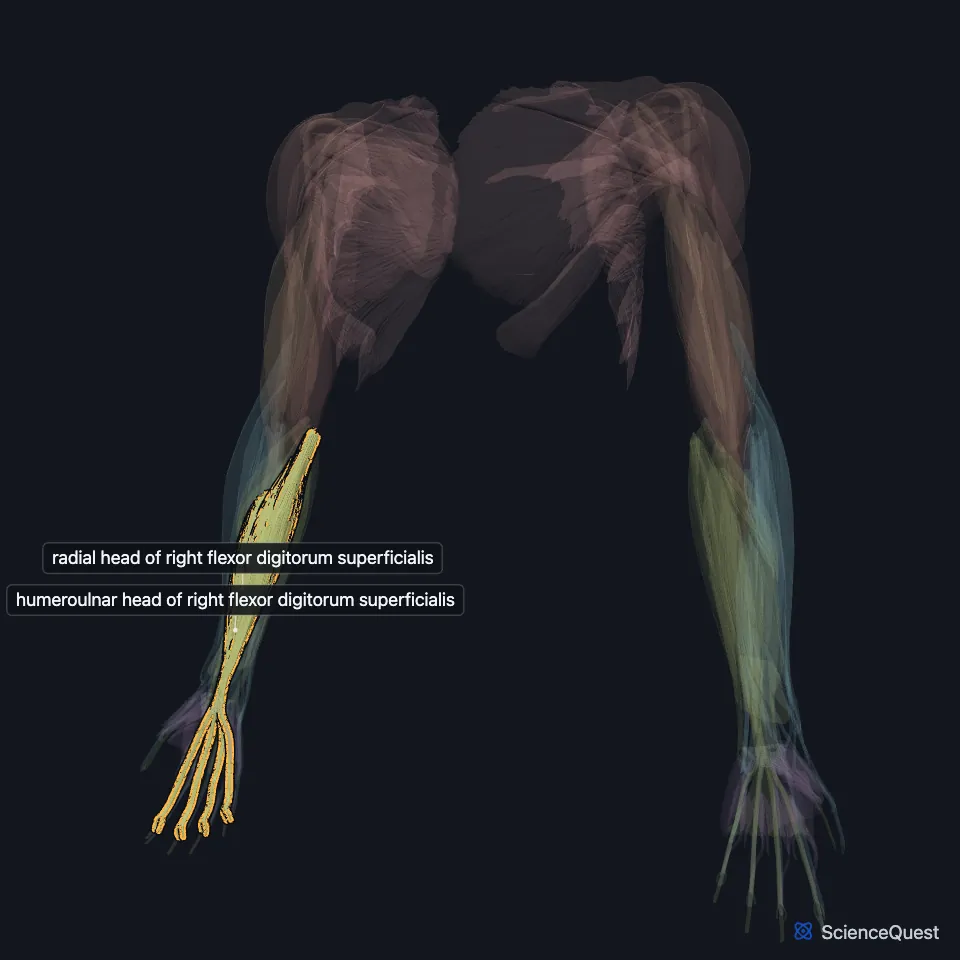
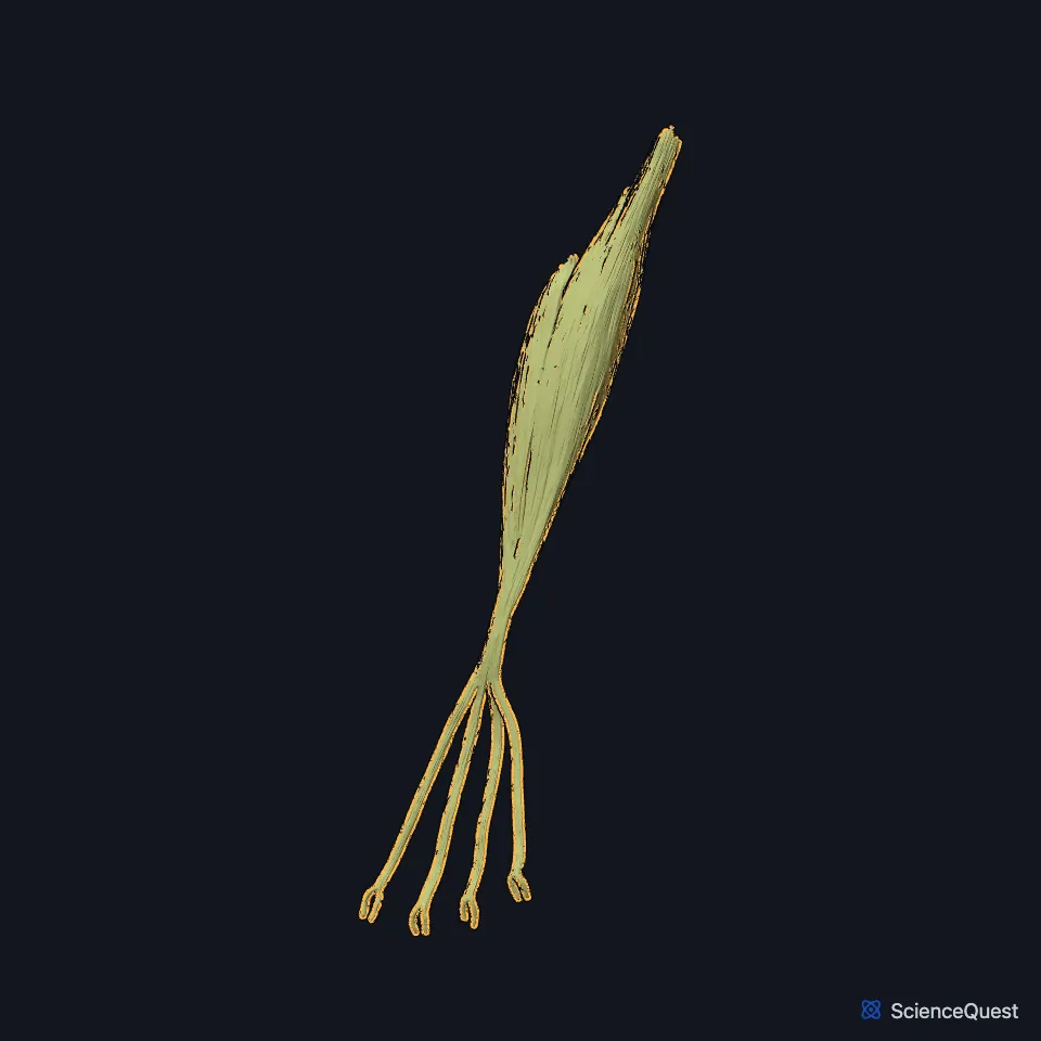
Open it in the Upper Limb Muscles Explorer See it alone in 3D
Where is the flexor digitorum superficialis?
It lies deep to the pronator teres, flexor carpi radialis, palmaris longus and flexor carpi ulnaris, and superficial to the flexor digitorum profundus and flexor pollicis longus. (StatPearls, Hand Flexor Digitorum Superficialis Muscle)
Its four tendons are four of the nine tendons in the carpal tunnel. They pass in pairs, the middle and ring finger tendons in front and the index and little finger tendons behind. (StatPearls, Hand Flexor Digitorum Superficialis Muscle; Gray’s Anatomy (1918), Volar Antibrachial Muscles)
What are the heads of the flexor digitorum superficialis?
StatPearls describes two heads. The humeroulnar head arises from the medial epicondyle and the coronoid process of the ulna, and the radial head from the upper half of the front of the radius. The median nerve and ulnar artery enter the forearm between them. (StatPearls, Hand Flexor Digitorum Superficialis Muscle)
Opposite the base of the proximal phalanx each tendon splits into two slips to let the profundus tendon through. The slips rejoin and insert on the middle phalanx. (Gray’s Anatomy (1918), Volar Antibrachial Muscles; StatPearls, Hand Flexor Digitorum Superficialis Muscle)
What does the flexor digitorum superficialis do?
It flexes the middle phalanges of the four fingers at the proximal interphalangeal joints, and it also flexes the knuckle (metacarpophalangeal) joints and the wrist. It can flex each finger on its own. (StatPearls, Hand Flexor Digitorum Superficialis Muscle)
To test it, the examiner holds three fingers straight, which takes the profundus out of action, and asks for the fourth to bend at its middle joint against resistance. (StatPearls, Hand Flexor Digitorum Superficialis Muscle)
Which nerve supplies the flexor digitorum superficialis?
The median nerve supplies it, from C7, C8 and T1. Weak flexion at the proximal interphalangeal joints is a sign of a high median neuropathy. (StatPearls, Hand Flexor Digitorum Superficialis Muscle and Median Nerve; Gray’s Anatomy (1918), Volar Antibrachial Muscles)
Its main blood supply comes from the ulnar artery. (StatPearls, Hand Flexor Digitorum Superficialis Muscle)
How does the flexor digitorum superficialis vary?
The part for the little finger can be missing from birth, which complicates the examination of a cut finger. In rare cases the muscle extends into the carpal tunnel and causes carpal tunnel symptoms. (StatPearls, Hand Flexor Digitorum Superficialis Muscle)
Where the sources differ
Sources disagree on the number of its heads. StatPearls describes two, humeroulnar and radial; Gray’s 1918 describes three, humeral, ulnar and radial, and calls the muscle flexor digitorum sublimis, a name StatPearls gives as former. (StatPearls, Hand Flexor Digitorum Superficialis Muscle; Gray’s Anatomy (1918), Volar Antibrachial Muscles)
They also place the insertion differently. Its own StatPearls article gives the bases of the middle phalanges, and the StatPearls median nerve article gives their sides; Gray’s 1918 has each tendon split again and insert on the sides of the second (middle) phalanx about its middle. (StatPearls, Hand Flexor Digitorum Superficialis Muscle and Median Nerve; Gray’s Anatomy (1918), Volar Antibrachial Muscles)
| Feature | Detail and source |
|---|---|
| Origin | Humeroulnar head: medial epicondyle and coronoid process of the ulna. Radial head: upper half of the front of the radius (StatPearls, Hand Flexor Digitorum Superficialis Muscle) |
| Insertion | Middle phalanges of the index, middle, ring and little fingers (StatPearls, Hand Flexor Digitorum Superficialis Muscle) |
| Action | Flexes the proximal interphalangeal joints; also the metacarpophalangeal joints and the wrist (StatPearls, Hand Flexor Digitorum Superficialis Muscle) |
| Nerve | Median nerve (C7, C8, T1) (StatPearls, Hand Flexor Digitorum Superficialis Muscle) |
Touches in this model
Measured on the BodyParts3D meshes: surfaces within 2 mm of the flexor digitorum superficialis, nearest first, 12 of 35.
- Flexor retinaculum of right wrist in contact
- Humeral head of right flexor carpi ulnaris in contact
- Humeral head of right pronator teres in contact
- Opponens digiti minimi of right hand in contact
- Right flexor carpi radialis in contact
- Right flexor digitorum profundus in contact
- Right flexor pollicis longus in contact
- Right palmaris longus in contact
- Right supinator in contact
- Set of lumbricals of right hand in contact
- Ulnar head of right pronator teres in contact
- Middle phalanx of right index finger in contact
Sources
Sources read
- StatPearls, Anatomy, Shoulder and Upper Limb, Hand Flexor Digitorum Superficialis Muscle. Lauren Okafor; Matthew A. Varacallo. Last update . CC BY-NC-ND 4.0. Read .
- Anatomy of the Human Body (Gray’s Anatomy), 20th US edition, 1918, Myology, The Muscles and Fasciae of the Forearm, 1. The Volar Antibrachial Muscles, pages 445 to 451; Internet Archive scan of the 1918 Lea and Febiger printing, terminology of 1918. Henry Gray; edited by Warren H. Lewis. Public domain. Read .
- StatPearls, Anatomy, Shoulder and Upper Limb, Median Nerve. Kelly A. Murphy; Nicholas Ryan; Adegbenro O. Fakoya. Last update . CC BY-NC-ND 4.0. Read .
The values are facts and free to use; this page’s selection and presentation are © 2026 ScienceQuest.