Flexor retinaculum of wrist
The flexor retinaculum of the wrist is a band of fibrous connective tissue that forms the roof of the carpal tunnel. Many experts treat it as the same structure as the transverse carpal ligament. (StatPearls, Wrist Flexor Retinaculum)
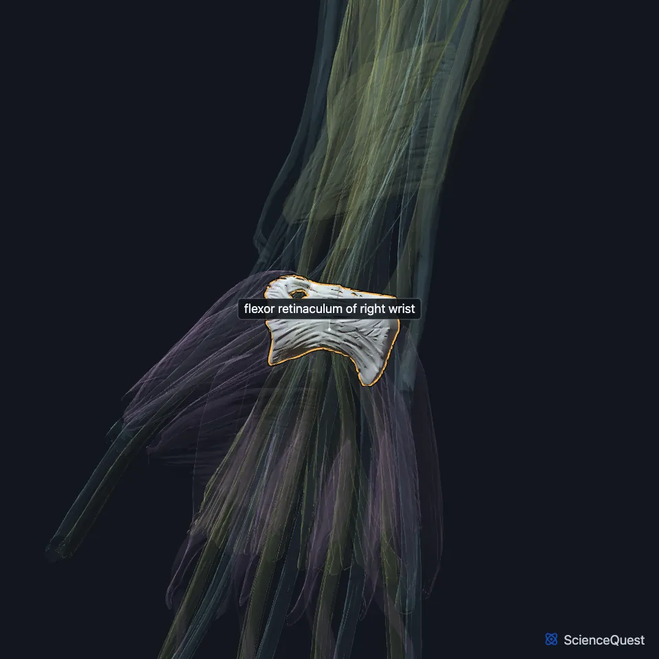
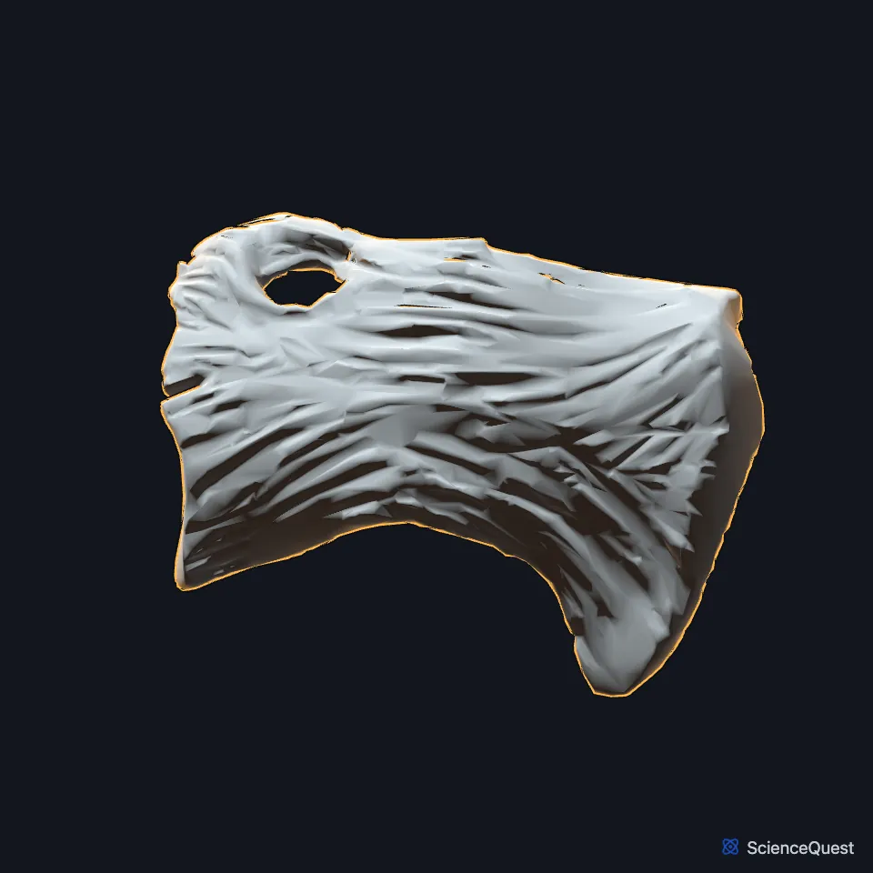
Open it in the Upper Limb Muscles Explorer See it alone in 3D
Where is the flexor retinaculum of the wrist?
It arches over the front of the carpal bones. On the radial side it attaches to the tuberosity of the scaphoid and the ridge of the trapezium, and on the ulnar side to the pisiform and the hook of the hamate. (StatPearls, Wrist Flexor Retinaculum; Gray’s Anatomy (1918), Transverse Carpal Ligament)
It is about 3 cm long and 2.5 cm wide, and it thickens from 1.5 mm to 6.0 mm toward the hand. (StatPearls, Wrist Flexor Retinaculum)
What is the flexor retinaculum of the wrist made of, and what joins it?
It begins where the lengthwise fibers of the forearm fascia thicken and turn crosswise. Its upper edge continues into the palmar carpal ligament and its lower edge merges with the palmar aponeurosis. (StatPearls, Wrist Flexor Retinaculum)
The palmaris longus tendon adheres to its front, and that muscle is absent in about 14% of people. Gray’s 1918, under the name transverse carpal ligament, says its lower border gives origin to the short muscles of the thumb and little finger. (StatPearls, Wrist Flexor Retinaculum; Gray’s Anatomy (1918), Transverse Carpal Ligament)
The FMA lists it as the flexor retinaculum of wrist, with flexor retinaculum of hand as a synonym and retinaculum musculorum flexorum manus as its Latin equivalent. (FMA, Flexor retinaculum of wrist)
What does the flexor retinaculum of the wrist do?
It covers nine tendons and a nerve: the four tendons of the flexor digitorum superficialis, the four of the flexor digitorum profundus, the tendon of the flexor pollicis longus and the median nerve. (StatPearls, Wrist Flexor Retinaculum)
It also works as a pulley for the carpal flexor muscles and helps hold the shape of the carpus. (StatPearls, Wrist Flexor Retinaculum)
What passes over and under the flexor retinaculum of the wrist?
The ulnar vessels and nerve cross in front of it, the nerve through the Guyon canal at its medial edge. The median nerve runs beneath it, and its recurrent branch leaves after the tunnel to supply the thenar muscles. (Gray’s Anatomy (1918), Transverse Carpal Ligament; StatPearls, Wrist Flexor Retinaculum)
Compression of the median nerve under it causes carpal tunnel syndrome, the most common compression neuropathy of the upper limb. If symptoms do not improve with conservative treatment, surgeons often cut or release the retinaculum to relieve the nerve. (StatPearls, Wrist Flexor Retinaculum)
The palmar cutaneous branch of the median nerve runs superficial to the carpal tunnel, so carpal tunnel syndrome spares the skin sensation over the thenar eminence. (StatPearls, Hand Thenar Eminence)
| Feature | Detail and source |
|---|---|
| Attached to | Radial side: scaphoid tuberosity and trapezial ridge; ulnar side: pisiform and hook of the hamate (StatPearls, Wrist Flexor Retinaculum) |
| Passes beneath it | Nine flexor tendons and the median nerve (StatPearls, Wrist Flexor Retinaculum) |
| Also called | Transverse carpal ligament (Gray’s 1918 name; many treat the two as one) (StatPearls, Wrist Flexor Retinaculum; Gray’s Anatomy (1918), Transverse Carpal Ligament) |
| Gives origin to | Short muscles of the thumb and little finger (Gray’s Anatomy (1918), Transverse Carpal Ligament) |
Touches in this model
Measured on the BodyParts3D meshes: surfaces within 2 mm of the flexor retinaculum of wrist, nearest first, 12 of 16.
- Abductor digiti minimi of right hand in contact
- Flexor digiti minimi brevis of right hand in contact
- Humeral head of right flexor carpi ulnaris in contact
- Humeroulnar head of right flexor digitorum superficialis in contact
- Opponens digiti minimi of right hand in contact
- Right abductor pollicis brevis in contact
- Right flexor carpi radialis in contact
- Right flexor pollicis longus in contact
- Right opponens pollicis in contact
- Right palmaris longus in contact
- Superficial head of right flexor pollicis brevis in contact
- Right pisiform in contact
Sources
Sources read
- StatPearls, Anatomy, Shoulder and Upper Limb, Wrist Flexor Retinaculum. Nathaniel Deak; Asa C. Black; Bruno Bordoni. Last update . CC BY-NC-ND 4.0. Read .
- Anatomy of the Human Body (Gray’s Anatomy), 20th US edition, 1918, Myology, The Muscles and Fasciae of the Hand, Transverse Carpal Ligament, pages 456 to 457; Internet Archive scan of the 1918 Lea and Febiger printing, terminology of 1918. Henry Gray; edited by Warren H. Lewis. Public domain. Read .
- Foundational Model of Anatomy, FMA39988, Flexor retinaculum of wrist (read on EBI OLS). FMA release terms, University of Washington. Read .
- StatPearls, Anatomy, Shoulder and Upper Limb, Hand Thenar Eminence. Dylan Luxenburg; Michael G. Rizzo. Last update . CC BY-NC-ND 4.0. Read .
The values are facts and free to use; this page’s selection and presentation are © 2026 ScienceQuest.