Extensor pollicis longus
The extensor pollicis longus is a deep muscle of the posterior forearm whose tendon makes a sharp turn at the Lister tubercle of the radius on its way to the thumb. It extends the thumb at the interphalangeal joint. (StatPearls, Forearm Muscles and Wrist Extensor Muscles)
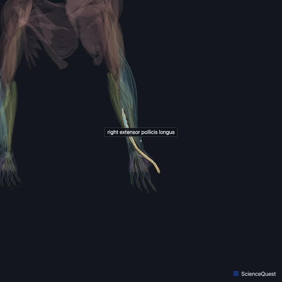
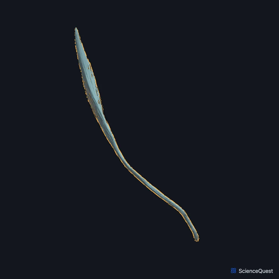
Open it in the Upper Limb Muscles Explorer See it alone in 3D
Where is the extensor pollicis longus?
It arises on the back of the middle third of the ulna, below the abductor pollicis longus, and from the interosseous membrane, then runs toward the radial side of the wrist. Its tendon is the only one in the third dorsal compartment. (Gray’s Anatomy (1918), Dorsal Antibrachial Muscles; StatPearls, Hand Extensor Pollicis Longus Muscle)
Beyond the retinaculum the tendon crosses the tendons of the two radial wrist extensors and forms the medial border of the anatomical snuffbox. The radial artery lies in the triangular gap between it and the extensor pollicis brevis tendon. (Gray’s Anatomy (1918), Dorsal Antibrachial Muscles; StatPearls, Hand Anatomical Snuff Box)
Where does the extensor pollicis longus insert?
It inserts on the base of the distal phalanx of the thumb. Over the thumb’s metacarpophalangeal joint a U-shaped sagittal band holds the tendon in place, and the radial part of that band matters more. (StatPearls, Wrist Extensor Muscles and Hand Extensor Pollicis Longus Muscle)
What does the extensor pollicis longus do?
It extends the thumb at the interphalangeal joint and the metacarpophalangeal joint. Its path through the third compartment also lets it help adduct the thumb. (StatPearls, Hand Extensor Pollicis Longus Muscle)
Studies find it produces more thumb extension force with the wrist extended 30° than with the wrist straight. (StatPearls, Hand Extensor Pollicis Longus Muscle)
Which nerve supplies the extensor pollicis longus, and why does its tendon rupture?
A motor branch of the posterior interosseous nerve, from its deep group of branches, supplies it. (StatPearls, Hand Extensor Pollicis Longus Muscle)
Its tendon is most vulnerable around the Lister tubercle, where it suffers mechanical wear and has a limited blood supply. It can rupture several weeks after a nondisplaced distal radius fracture, in up to 5% of adults with that fracture. (StatPearls, Wrist Extensor Muscles and Hand Extensor Pollicis Longus Muscle)
Surgeons most often repair a ruptured tendon by transferring the extensor indicis tendon to it. (StatPearls, Wrist Extensor Muscles)
How does the extensor pollicis longus vary?
Variants occur in about 1% of people. They include doubled tendons and tendon links to the index finger extensors, and they rarely cause symptoms. (StatPearls, Hand Extensor Pollicis Longus Muscle)
| Feature | Detail and source |
|---|---|
| Origin | Back of the middle third of the ulna; interosseous membrane (Gray’s Anatomy (1918), Dorsal Antibrachial Muscles; StatPearls, Hand Extensor Pollicis Longus Muscle) |
| Insertion | Base of the distal phalanx of the thumb (StatPearls, Wrist Extensor Muscles) |
| Action | Extends the thumb at the interphalangeal and metacarpophalangeal joints; assists thumb adduction (StatPearls, Hand Extensor Pollicis Longus Muscle) |
| Nerve | Posterior interosseous nerve (StatPearls, Hand Extensor Pollicis Longus Muscle) |
Touches in this model
Measured on the BodyParts3D meshes: surfaces within 2 mm of the extensor pollicis longus, nearest first, 12 of 19.
- Right abductor pollicis longus in contact
- Right extensor carpi radialis brevis in contact
- Right extensor carpi radialis longus in contact
- Right extensor digitorum in contact
- Right extensor indicis in contact
- Right extensor pollicis brevis in contact
- Right flexor digitorum profundus in contact
- Ulnar head of right extensor carpi ulnaris in contact
- Distal phalanx of right thumb in contact
- Interosseous membrane of right forearm in contact
- Proximal phalanx of right thumb in contact
- Right first metacarpal bone in contact
Sources
Sources read
- StatPearls, Anatomy, Shoulder and Upper Limb, Forearm Muscles. Brittney Mitchell; Lacey Whited. Last update . CC BY-NC-ND 4.0. Read .
- StatPearls, Anatomy, Shoulder and Upper Limb, Wrist Extensor Muscles. Nicole Reeves; Tyler Crow; Matthew A. Varacallo. Last update . CC BY-NC-ND 4.0. Read .
- Anatomy of the Human Body (Gray’s Anatomy), 20th US edition, 1918, Myology, The Muscles and Fasciae of the Forearm, 2. The Dorsal Antibrachial Muscles, pages 451 to 456; Internet Archive scan of the 1918 Lea and Febiger printing, terminology of 1918. Henry Gray; edited by Warren H. Lewis. Public domain. Read .
- StatPearls, Anatomy, Shoulder and Upper Limb, Hand Extensor Pollicis Longus Muscle. Irfan A. Khan; Marjorie V. Launico. Last update . CC BY-NC-ND 4.0. Read .
- StatPearls, Anatomy, Shoulder and Upper Limb: Hand Anatomical Snuff Box. Sasha Hallett Reid; Felix Jozsa; John V. Ashurst. Last update . CC BY-NC-ND 4.0. Read .
The values are facts and free to use; this page’s selection and presentation are © 2026 ScienceQuest.