Papillary muscle of left ventricle
The left ventricle classically has two papillary muscles, the anterolateral and the posteromedial. Through the chordae tendineae they hold the two leaflets of the mitral valve. (StatPearls, Heart Papillary Muscles and Mitral Valve)
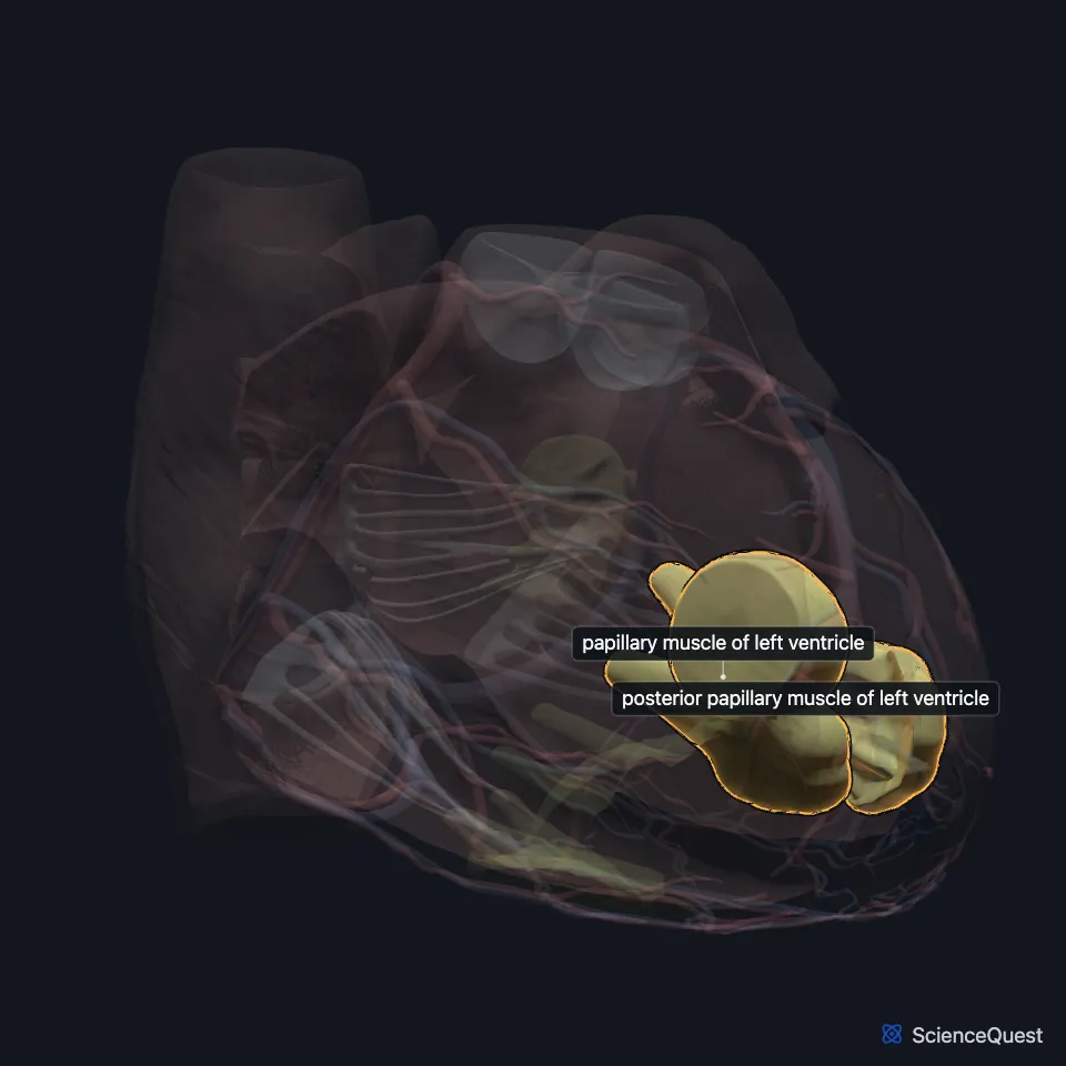
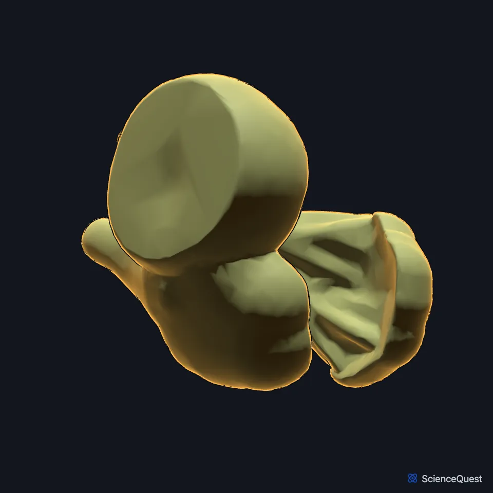
Where are the papillary muscles of the left ventricle?
The anterolateral muscle arises from the sternocostal wall of the ventricle and the posteromedial muscle from the diaphragmatic wall. (StatPearls, Heart Papillary Muscles)
StatPearls (Mitral Valve) adds that both start from the apical part of the ventricle and are named for their position relative to the mitral commissures. (StatPearls, Mitral Valve)
What do the papillary muscles of the left ventricle do?
Each muscle sends chordae to both mitral leaflets. As the ventricle contracts in systole, the muscles pull the chordae taut and keep the leaflets from prolapsing or turning back into the left atrium. (StatPearls, Mitral Valve and Heart Papillary Muscles; Gray 1918, 4b. The Heart)
When cardiomyopathy or ischemia remodels the ventricle, the change in its size pulls the papillary muscles out of place. The leaflets then no longer meet, and the valve leaks: secondary, or functional, mitral regurgitation. (StatPearls, Heart Papillary Muscles)
What lies next to the papillary muscles of the left ventricle?
Gray’s Anatomy (1918) attaches one to the anterior and one to the posterior wall, among trabeculae carneae that are more numerous than on the right and densely interlaced, especially at the apex and on the posterior wall. (Gray 1918, 4b. The Heart)
With the annulus, the two leaflets and the chordae, they make up the mitral valve apparatus between the left atrium and the left ventricle. (StatPearls, Mitral Valve)
Where do the left ventricle’s papillary muscles get their blood?
The anterolateral muscle is fed by branches of the left coronary artery, the anterior interventricular and circumflex arteries, and more often has more than one supplying vessel. (StatPearls, Heart Papillary Muscles and Mitral Valve)
The posteromedial muscle most often depends on the right coronary artery, sometimes on the circumflex. In one study it had a single supplying vessel in 63% of patients. (StatPearls, Heart Papillary Muscles)
Why does the posteromedial papillary muscle rupture more often?
Its usual single supply from the posterior descending artery makes it rupture 6 to 12 times more often than the anterolateral muscle, and it can follow an inferior wall infarction. (StatPearls, Papillary Muscle Rupture and Heart Papillary Muscles)
Rupture usually comes 2 to 7 days after the infarction and causes severe acute mitral regurgitation, cardiogenic shock and pulmonary edema. It needs prompt surgery, usually mitral valve repair or replacement. (StatPearls, Papillary Muscle Rupture)
How do the left ventricle’s papillary muscles vary?
Their shape varies from conical to bifurcated or trifurcated, and each carries 2 to 20 chordae. In a parachute mitral valve all the chordae attach to a single papillary muscle, which often narrows the valve. (StatPearls, Heart Papillary Muscles)
Where the sources differ
The sources name the two muscles differently. StatPearls (Heart Papillary Muscles) uses anterolateral and posteromedial, and StatPearls (Mitral Valve) uses both pairs. Gray’s Anatomy (1918) ties one to the anterior wall and one to the posterior, and its account of the heart’s muscle fibers, which follows McCallum, calls them the anterior and posterior papillary muscles. (StatPearls, Heart Papillary Muscles and Mitral Valve; Gray 1918, 4b. The Heart)
| Feature | Detail and source |
|---|---|
| Number | Two (StatPearls, Heart Papillary Muscles; Gray 1918, 4b. The Heart) |
| Arise from | Sternocostal wall (anterolateral) and diaphragmatic wall (posteromedial) (StatPearls, Heart Papillary Muscles) |
| Holds | Mitral valve, both leaflets from each muscle (StatPearls, Mitral Valve) |
Touches in this model
Measured on the BodyParts3D meshes: surfaces within 2 mm of the papillary muscle of left ventricle, nearest first.
- Mitral valve in contact
- Set of interventricular septal branches of left coronary artery in contact
- Wall of heart in contact
Sources
Sources read
- StatPearls, Anatomy, Thorax, Heart Papillary Muscles. Natalie L. Rich; Yusuf S. Khan. Last update . CC BY-NC-ND 4.0. Read .
- StatPearls, Anatomy, Thorax, Mitral Valve. Felipe Sanchez Vaca; Bruno Bordoni. Last update . CC BY-NC-ND 4.0. Read .
- Gray’s Anatomy of the Human Body, 20th US edition (1918), V. Angiology, 4b. The Heart (1918 terms: longitudinal sulci for interventricular sulci, musculi papillares for papillary muscles). Henry Gray. Public domain. Read .
- StatPearls, Papillary Muscle Rupture. Sanjeev Sharma; Lauren V. Burton; Kevin Beier. Last update . CC BY-NC-ND 4.0. Read .
The values are facts and free to use; this page’s selection and presentation are © 2026 ScienceQuest.