Papillary muscle of right ventricle
The papillary muscles of the right ventricle are pillar-like muscles that project from the ventricle’s inner wall. Through the chordae tendineae they attach to the leaflets of the tricuspid valve. (StatPearls, Heart Papillary Muscles; Gray 1918, 4b. The Heart)
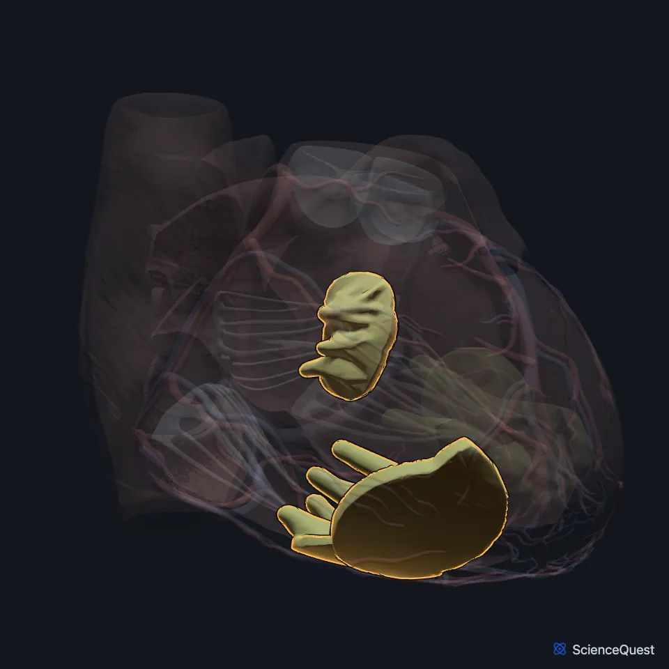
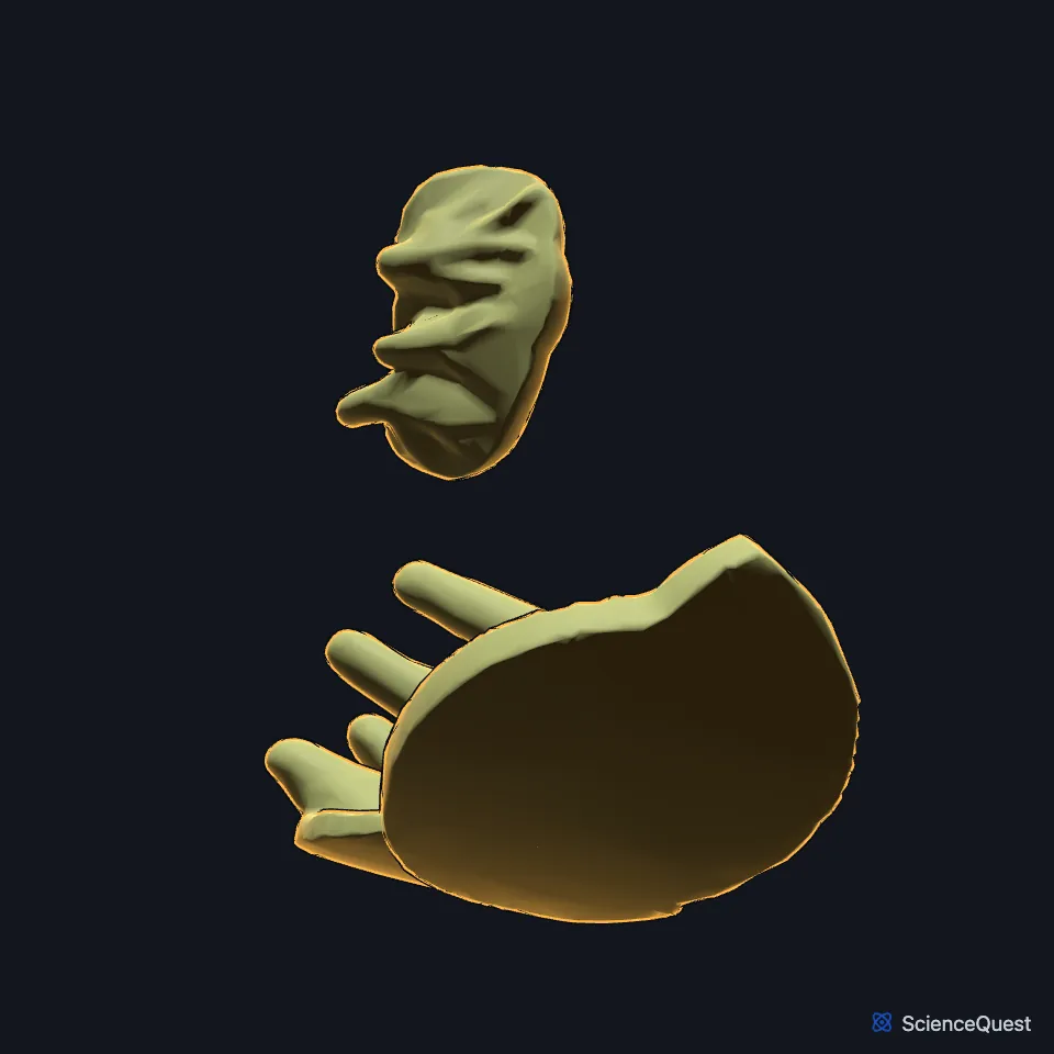
Where are the papillary muscles of the right ventricle?
StatPearls (Heart Papillary Muscles) describes three. The anterior is the largest and arises from the anterior wall, the posterior from the inferior wall, and the septal, the smallest, from the interventricular septum. (StatPearls, Heart Papillary Muscles)
What do the papillary muscles of the right ventricle do?
When the ventricles contract in systole, the papillary muscles contract with them and pull the chordae tight. This stops the tricuspid cusps from prolapsing or turning inside out into the right atrium. (StatPearls, Heart Papillary Muscles; Gray 1918, 4b. The Heart)
The right bundle branch splits into Purkinje fibers at the base of the papillary muscles. In Gray’s Anatomy (1918) its strands end in the papillary muscles and in the ventricular muscle generally. (StatPearls, Heart Papillary Muscles; Gray 1918, 4b. The Heart)
Which tricuspid cusps does each muscle hold?
The anterior muscle’s chordae hold the anterior and posterior leaflets, and the posterior muscle’s chordae the posterior and septal leaflets. Gray’s Anatomy (1918) calls the septal leaflet the medial cusp. The posterior muscle can have two or three parts. (StatPearls, Tricuspid Valve Repair; FMA, FMA7240; Gray 1918, 4b. The Heart)
The septomarginal trabecula, or moderator band, is a muscle band running from the septum to the anterior papillary muscle. Gray’s Anatomy (1918) finds it frequently and StatPearls (Tricuspid Valve Repair) says it sometimes joins this muscle. Gray explains the name moderator band by a possible role in keeping the ventricle from overstretching. (FMA, FMA7272; Gray 1918, 4b. The Heart; StatPearls, Tricuspid Valve Repair)
What lies next to the papillary muscles of the right ventricle?
They are one of three kinds of trabeculae carneae, the muscular columns on the inner surface of the ventricle: the kind joined to the wall of the heart by their bases, with chordae arising from their tips. (Gray 1918, 4b. The Heart)
The chordae tendineae are thin fibrous cords linking the papillary muscles to the valve leaflets. (StatPearls, Heart Papillary Muscles)
Where do the right ventricle’s papillary muscles get their blood?
The anterior muscle is most often supplied by branches of both the left coronary artery and the right coronary artery, and in some people by the left alone. Most papillary muscles draw on more than one artery, with wide variation. (StatPearls, Heart Papillary Muscles)
Can the right ventricle’s papillary muscles rupture?
Rupture of the tricuspid papillary muscles can follow myocardial ischemia, trauma or infective endocarditis. Blood then leaks back through the valve, which can lead to right-sided heart failure. (StatPearls, Papillary Muscle Rupture)
Where the sources differ
The sources count them differently, and StatPearls disagrees with itself. Its Heart Papillary Muscles article describes three, anterior, posterior and septal; its Tricuspid Valve Repair article usually finds two, anterior and posterior, with a variable third septal muscle. Gray’s Anatomy (1918) describes two, with further chordae arising straight from the septum or from small papillary eminences on it. (StatPearls, Heart Papillary Muscles and Tricuspid Valve Repair; Gray 1918, 4b. The Heart)
| Feature | Detail and source |
|---|---|
| Number | Three in StatPearls (Heart Papillary Muscles); usually two plus a variable septal one in StatPearls (Tricuspid Valve Repair); two in Gray’s Anatomy (1918) (StatPearls, Heart Papillary Muscles and Tricuspid Valve Repair; Gray 1918, 4b. The Heart) |
| Largest | Anterior papillary muscle (StatPearls, Heart Papillary Muscles; Gray 1918, 4b. The Heart) |
| Holds | Tricuspid valve, through the chordae tendineae (StatPearls, Heart Papillary Muscles) |
Touches in this model
Measured on the BodyParts3D meshes: surfaces within 2 mm of the papillary muscle of right ventricle, nearest first.
- Anterior interventricular branch of left coronary artery in contact
- Great cardiac vein in contact
- Marginal branch of right coronary artery in contact
- Set of anterior cardiac veins in contact
- Tricuspid valve in contact
- Wall of heart in contact
- Posterior interventricular branch of right coronary artery 0.3 mm away
- Set of interventricular septal branches of right coronary artery 0.5 mm away
- Diaphragm 0.5 mm away
- Middle cardiac vein 0.6 mm away
Sources
Sources read between and
- StatPearls, Anatomy, Thorax, Heart Papillary Muscles. Natalie L. Rich; Yusuf S. Khan. Last update . CC BY-NC-ND 4.0. Read .
- Gray’s Anatomy of the Human Body, 20th US edition (1918), V. Angiology, 4b. The Heart (1918 terms: longitudinal sulci for interventricular sulci, musculi papillares for papillary muscles). Henry Gray. Public domain. Read .
- StatPearls, Tricuspid Valve Repair. Sohail K. Mahboobi; Sanjeev Sharma; Andaleeb A. Ahmed. Last update . CC BY-NC-ND 4.0. Read .
- Foundational Model of Anatomy, via the EMBL-EBI Ontology Lookup Service, Septal leaflet of tricuspid valve (FMA7240), preferred name and definition. Not printed on the page. Read .
- Foundational Model of Anatomy, via the EMBL-EBI Ontology Lookup Service, Septomarginal trabecula (FMA7272), preferred name, synonyms and definition. Not printed on the page. Read .
- StatPearls, Papillary Muscle Rupture. Sanjeev Sharma; Lauren V. Burton; Kevin Beier. Last update . CC BY-NC-ND 4.0. Read .
The values are facts and free to use; this page’s selection and presentation are © 2026 ScienceQuest.