Central canal of spinal cord
The central canal is a fine channel of cerebrospinal fluid that runs down the length of the spinal cord, in the middle of its gray matter. At its upper end it opens into the fourth ventricle. (Gray 1918, 3. The Spinal Cord; StatPearls, Spinal Cord Morphology)
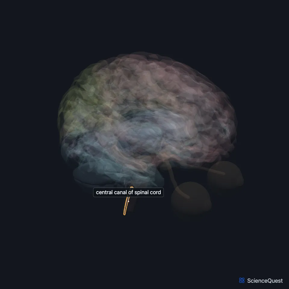
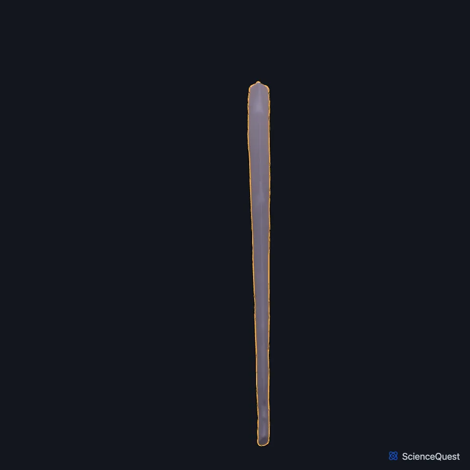
Where is the central canal?
It runs through the gray commissure that joins the two halves of the H-shaped gray matter, and continues upward through the lower medulla oblongata. (StatPearls, Spinal Cord; Gray 1918, 3. The Spinal Cord)
Gray’s 1918 places it in the front third of the cord in the neck and chest, near the middle in the lumbar enlargement, and near the back surface in the conus medullaris. (Gray 1918, 3. The Spinal Cord)
What lines the central canal?
It is lined by ependymal cells and filled with cerebrospinal fluid. The gray matter around it is lamina X, the central zone of the spinal laminae. (StatPearls, Spinal Cord and Spinal Cord Morphology; Gray 1918, 3. The Spinal Cord)
It is what is left of the cavity of the embryo’s neural tube, which narrows down to form it. (StatPearls, Spinal Cord)
What does the central canal do?
It carries the ventricular system on below the brain: the ventricles are continuous with it, and part of the cerebrospinal fluid flows down into it. (StatPearls, Ventricular System and Hydrocephalus)
What lies around the central canal?
In front of it lie the anterior gray commissure and then the anterior white commissure, and behind it the posterior gray commissure. At its lower end it widens into the terminal ventricle, 8 to 10 mm long, in the conus medullaris. (Gray 1918, 3. The Spinal Cord; StatPearls, Spinal Cord)
What is syringomyelia?
Syringomyelia is a fluid-filled cavity, a syrinx, in the cord tissue or the central canal, often with a Chiari type 1 malformation. In the communicating form the dilated canal connects with the fourth ventricle and comes with hydrocephalus. (StatPearls, Syringomyelia)
The canal may close off with age, and a canal that stays open is not an anomaly. Gray’s 1918 notes that the terminal ventricle tends to close after about age 40. (StatPearls, Syringomyelia; Gray 1918, 3. The Spinal Cord)
Touches in this model
Measured on the BodyParts3D meshes: surfaces within 2 mm of the central canal of spinal cord, nearest first.
- Fourth ventricle in contact
- Medulla oblongata in contact
Sources
Sources read
- Gray’s Anatomy of the Human Body, 20th US edition (1918), IX. Neurology, 3. The Spinal Cord or Medulla Spinalis. Henry Gray. Public domain. Read .
- StatPearls, Neuroanatomy, Spinal Cord Morphology. Muthu Kuzhali Ganapathy; Vamsi Reddy; Prasanna Tadi. Last update . CC BY-NC-ND 4.0. Read .
- StatPearls, Neuroanatomy, Spinal Cord. Yusuf S. Khan; Forshing Lui. Last update . CC BY-NC-ND 4.0. Read .
- StatPearls, Neuroanatomy, Ventricular System. Saraswati Satyanarayan Shenoy; Forshing Lui. Last update . CC BY-NC-ND 4.0. Read .
- StatPearls, Hydrocephalus. Miroslava Koleva; Orlando De Jesus. Last update . CC BY-NC-ND 4.0. Read .
- StatPearls, Syringomyelia. Varadaraya Satyanarayan Shenoy; Sunil Munakomi; Raghuram Sampath. Last update . CC BY-NC-ND 4.0. Read .
The values are facts and free to use; this page’s selection and presentation are © 2026 ScienceQuest.