Cerebral aqueduct
The cerebral aqueduct, or aqueduct of Sylvius, is the narrow channel through the midbrain that carries cerebrospinal fluid from the third ventricle to the fourth ventricle. It is the narrowest part of the ventricular system. (StatPearls, Cerebral Aqueduct (Sylvian); Gray 1918, 4b. The Mid-brain)
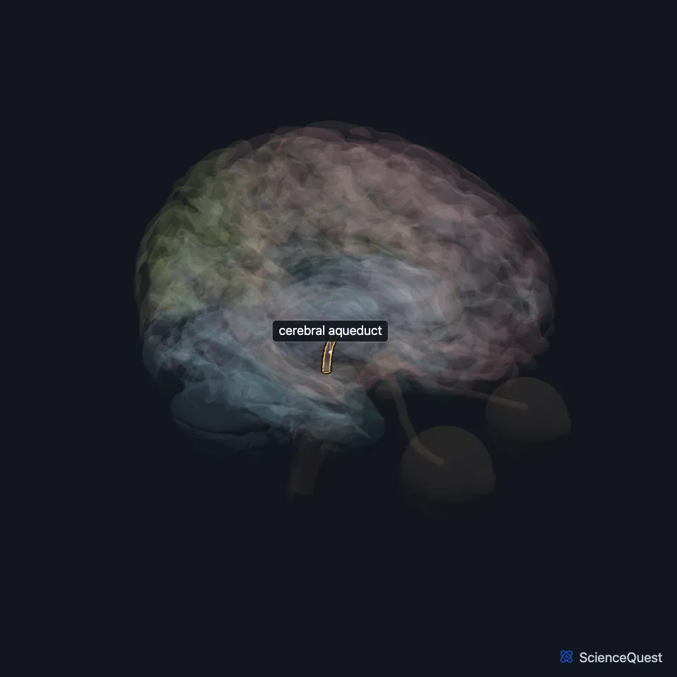
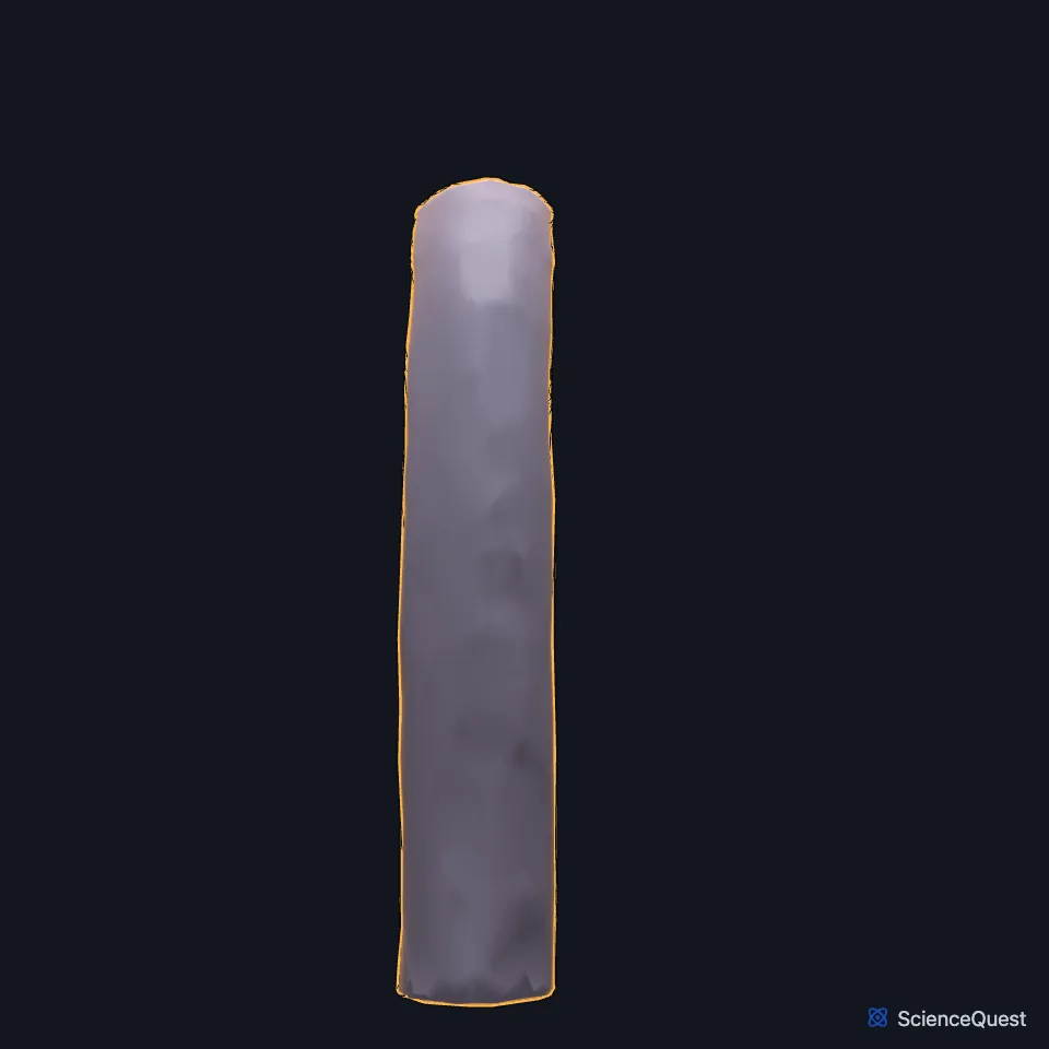
Where is the cerebral aqueduct?
It runs between the colliculi at the back of the midbrain and the tegmentum in front. (Gray 1918, 4b. The Mid-brain; StatPearls, Mesencephalon Midbrain)
It begins under the posterior commissure. Its middle part, the antrum, lies between two narrowings, at the superior colliculus and at the inferior colliculus, and its last part, the narrowest, opens into the fourth ventricle. (StatPearls, Cerebral Aqueduct (Sylvian))
What lies around the cerebral aqueduct?
A ring of gray matter, the periaqueductal gray, surrounds it. The nuclei of the oculomotor and trochlear nerves lie in the front part of that gray matter, and the pineal body sits close by, so a pineal tumor can press on the channel. (StatPearls, Brainstem and Cerebral Aqueduct (Sylvian); Gray 1918, 4b. The Mid-brain)
What lines the cerebral aqueduct, and how does it form?
Like the rest of the ventricular system, it is lined with ependymal cells. Gray’s 1918 describes this lining as ciliated columnar epithelium. (StatPearls, Cerebral Aqueduct (Sylvian); Gray 1918, 4b. The Mid-brain)
It forms from the cavity of the embryo’s midbrain vesicle, which narrows, and it keeps narrowing until about ten weeks after birth. (StatPearls, Cerebral Aqueduct (Sylvian))
What does the cerebral aqueduct do?
Fluid made by the choroid plexus of the lateral and third ventricles passes through it to reach the fourth ventricle, and speeds up as the channel narrows toward its lower end. It has no choroid plexus of its own. (StatPearls, Hydrocephalus, Cerebral Aqueduct (Sylvian) and Choroid Plexus)
What happens when the cerebral aqueduct narrows?
Aqueductal stenosis is a common cause of hydrocephalus, with one peak before age 1 and another after age 12. Almost 10% of hydrocephalus in newborns comes from a brain stem malformation with a narrowed aqueduct. (StatPearls, Cerebral Aqueduct (Sylvian) and Hydrocephalus)
Tectal plate gliomas and pineal tumors can press it shut. Treatment for the resulting hydrocephalus is a ventriculoperitoneal shunt or an endoscopic third ventriculostomy. (StatPearls, Cerebral Aqueduct (Sylvian))
Where the sources differ
Its length is given as about 15 mm by Gray’s 1918 and by StatPearls’ aqueduct article, but as approximately 18 mm by StatPearls’ article on the ventricular system. (Gray 1918, 4b. The Mid-brain; StatPearls, Cerebral Aqueduct (Sylvian) and Ventricular System)
Touches in this model
Measured on the BodyParts3D meshes: surfaces within 2 mm of the cerebral aqueduct, nearest first.
- Fourth ventricle in contact
- Midbrain in contact
- Pons in contact
- Third ventricle in contact
- Posterior commissure 0.5 mm away
- Left inferior colliculus 0.6 mm away
- Right inferior colliculus 0.6 mm away
- Right superior colliculus 1.5 mm away
- Left superior colliculus 1.6 mm away
Sources
Sources read
- StatPearls, Neuroanatomy, Cerebral Aqueduct (Sylvian). Jessica M. Rubino; Jeffery P. Hogg. Last update . CC BY-NC-ND 4.0. Read .
- Gray’s Anatomy of the Human Body, 20th US edition (1918), IX. Neurology, 4b. The Mid-brain or Mesencephalon. Henry Gray. Public domain. Read .
- StatPearls, Neuroanatomy, Mesencephalon Midbrain. Francheska Caminero; Marco Cascella. Last update . CC BY-NC-ND 4.0. Read .
- StatPearls, Neuroanatomy, Brainstem. Hayden Basinger; Jeffery P. Hogg. Last update . CC BY-NC-ND 4.0. Read .
- StatPearls, Hydrocephalus. Miroslava Koleva; Orlando De Jesus. Last update . CC BY-NC-ND 4.0. Read .
- StatPearls, Neuroanatomy, Choroid Plexus. Kinaan Javed; Vamsi Reddy; Forshing Lui. Last update . CC BY-NC-ND 4.0. Read .
- StatPearls, Neuroanatomy, Ventricular System. Saraswati Satyanarayan Shenoy; Forshing Lui. Last update . CC BY-NC-ND 4.0. Read .
The values are facts and free to use; this page’s selection and presentation are © 2026 ScienceQuest.