Superior colliculus
The superior colliculi are a pair of rounded bumps on the back of the upper midbrain. They take in what the eyes see, combine it with sound and touch, and help steer the eyes and head toward or away from what was sensed. (StatPearls, Superior Colliculus; Gray 1918, 4b. The Mid-brain)
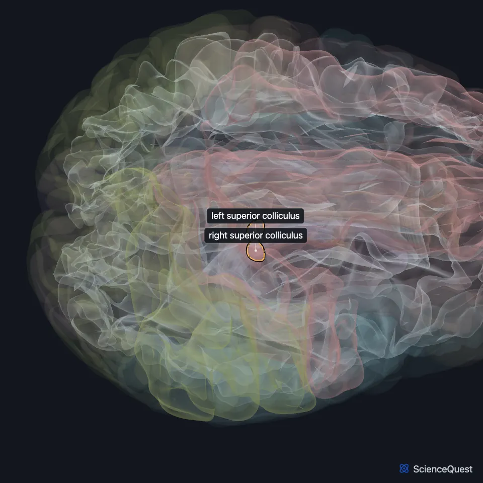
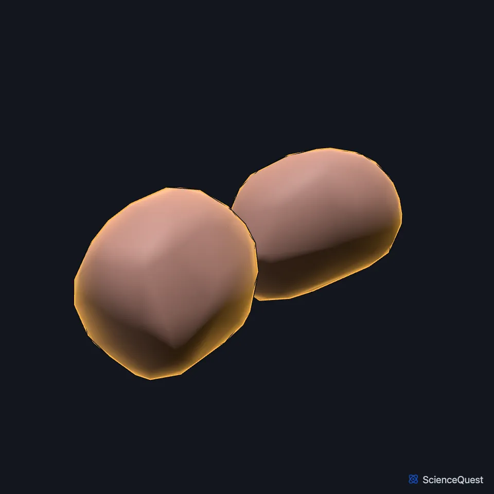
Where is the superior colliculus?
It sits on the back of the midbrain, above the inferior colliculus and below the pineal body. The four colliculi together form the roof of the midbrain, the tectum, which Gray’s 1918 calls the corpora quadrigemina. (StatPearls, Superior Colliculus, Mesencephalon Midbrain and Inferior Colliculus; Gray 1918, 4b. The Mid-brain)
How is the superior colliculus built?
It has seven cell layers in three groups. The superficial layers respond only to vision, while the intermediate and deep layers also take in hearing and touch. (StatPearls, Superior Colliculus)
From each colliculus a band of fibers, the superior brachium, runs forward to the lateral geniculate body and the optic tract. (StatPearls, Brainstem; Gray 1918, 4b. The Mid-brain)
What does the superior colliculus do?
It helps turn the head and eyes toward or away from a stimulus, and when the head cannot move it takes part in the quick eye jumps called saccades. Its deeper layers also pick up a looming object, such as an approaching snake, and pass the warning on toward defensive behavior. (StatPearls, Superior Colliculus)
What connects to the superior colliculus?
Most of its visual input comes straight from the retina along the optic nerve and optic tract, with less from the visual cortex. Sound reaches it from the inferior colliculi. (StatPearls, Superior Colliculus; Neuroscience Online, Visual Processing: Cortical Pathways)
Its output reaches the nuclei of the three nerves that move the eyes, the oculomotor, trochlear and abducens, so all six of the eye’s moving muscles are under its influence during a gaze shift. (StatPearls, Superior Colliculus)
What happens when the superior colliculus is damaged?
A tectal glioma, a rare low-grade tumor mostly of children, involves both pairs of colliculi and the cerebral aqueduct, and raises the pressure inside the skull. Surgery here risks Parinaud syndrome, whose hallmark is weak upward gaze. (StatPearls, Superior Colliculus and Parinaud Syndrome)
Where the sources differ
StatPearls counts seven layers in superficial, intermediate and deep groups, while Gray’s 1918 describes four strata, from the stratum zonale on the surface to the deep stratum lemnisci. (StatPearls, Superior Colliculus; Gray 1918, 4b. The Mid-brain)
Touches in this model
Measured on the BodyParts3D meshes: surfaces within 2 mm of the superior colliculus, nearest first, 12 of 13.
- Brachium of left superior colliculus in contact
- Brachium of right superior colliculus in contact
- Midbrain in contact
- Brachium of right inferior colliculus 0.2 mm away
- Brachium of left inferior colliculus 0.3 mm away
- Posterior commissure 0.5 mm away
- Left inferior colliculus 1.0 mm away
- Right inferior colliculus 1.0 mm away
- Cerebral aqueduct 1.5 mm away
- Left thalamus 1.6 mm away
- Right thalamus 1.7 mm away
- Pineal body 1.8 mm away
Sources
Sources read
- StatPearls, Neuroanatomy, Superior Colliculus. Ryan D. Zubricky; Joe M. Das. Last update . CC BY-NC-ND 4.0. Read .
- Gray’s Anatomy of the Human Body, 20th US edition (1918), IX. Neurology, 4b. The Mid-brain or Mesencephalon. Henry Gray. Public domain. Read .
- StatPearls, Neuroanatomy, Mesencephalon Midbrain. Francheska Caminero; Marco Cascella. Last update . CC BY-NC-ND 4.0. Read .
- StatPearls, Neuroanatomy, Inferior Colliculus. Margaret E. Driscoll; Prasanna Tadi. Last update . CC BY-NC-ND 4.0. Read .
- StatPearls, Neuroanatomy, Brainstem. Hayden Basinger; Jeffery P. Hogg. Last update . CC BY-NC-ND 4.0. Read .
- Neuroscience Online, McGovern Medical School, Section 2, Chapter 15, Visual Processing: Cortical Pathways. Valentin Dragoi, Ph.D. (content provided by Chieyeko Tsuchitani, Ph.D.). Reviewed . Open access; no reuse licence stated. Read .
- StatPearls, Parinaud Syndrome. Kaberi B. Feroze; Bhupendra C. Patel. Last update . CC BY-NC-ND 4.0. Read .
The values are facts and free to use; this page’s selection and presentation are © 2026 ScienceQuest.