Optic tract
Each optic tract is a bundle of nerve fibers that carries visual signals from the optic chiasm back to the lateral geniculate body. Each tract carries one half of the visual field: the left tract the right half, and the right tract the left half. (StatPearls, Optic Tract)
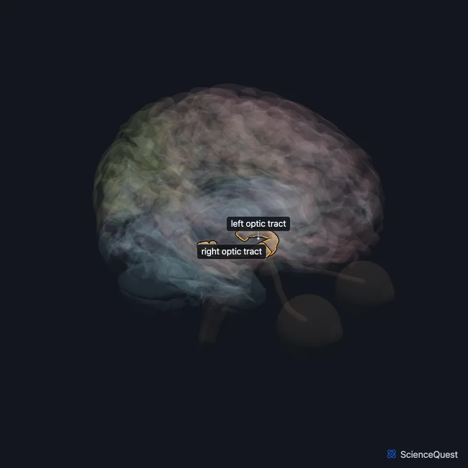
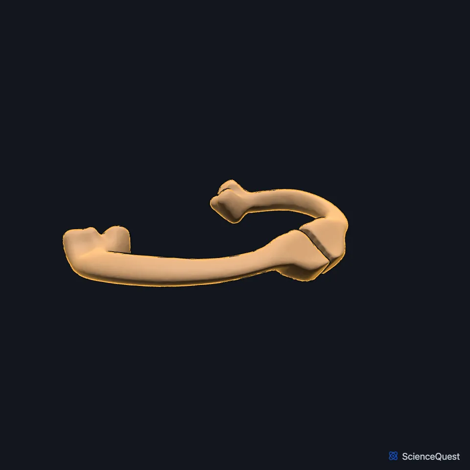
Where does the optic tract run?
From the chiasm it runs back and outward around the side of the cerebral peduncle to the lateral geniculate body. On the way the pituitary stalk and the cerebral peduncles lie on its inner side. (StatPearls, Optic Tract; Gray 1918, 4c. The Fore-brain)
Which fibers does the optic tract carry?
It is made of the axons of retinal ganglion cells. Each tract holds fibers from the temporal half of the retina on its own side and from the nasal half of the opposite retina, which crossed at the chiasm. (StatPearls, Optic Tract; Neuroscience Online, Visual Processing: Cortical Pathways)
At the chiasm the crossed and uncrossed fibers are about 53 to 47. (StatPearls, Optic Tract)
Where do optic tract fibers end?
Most end in the lateral geniculate body, the relay for seeing. Others go to the superior colliculus for eye movements, to the pretectum of the midbrain for the pupillary light reflex, and to the hypothalamus. (Neuroscience Online, Visual Processing: Cortical Pathways; StatPearls, Optic Tract)
What supplies the optic tract?
Its blood comes from branches of the posterior communicating artery and the anterior choroidal artery. The anterior choroidal artery runs beside the tract and then on to the lateral geniculate body. (StatPearls, Optic Tract)
What happens when an optic tract is damaged?
Damage to one optic tract most often causes a homonymous hemianopia, loss of the opposite half of the visual field in both eyes. A left tract lesion, for example, takes away the right half of what each eye sees. (StatPearls, Optic Tract)
Stroke is the most common cause of homonymous field defects. Surgeons removing the front of the temporal lobe for epilepsy have to watch for the tract. (StatPearls, Optic Tract)
Touches in this model
Measured on the BodyParts3D meshes: surfaces within 2 mm of the optic tract, nearest first, 12 of 33.
- Brachium of left inferior colliculus in contact
- Brachium of right inferior colliculus in contact
- Hypothalamus in contact
- Lamina terminalis in contact
- Left amygdala in contact
- Left lateral geniculate body in contact
- Left medial geniculate body in contact
- Left optic nerve in contact
- Left stria terminalis in contact
- Optic chiasm in contact
- Peduncle of midbrain in contact
- Right amygdala in contact
Sources
Sources read
- StatPearls, Neuroanatomy, Optic Tract. Divy Mehra; Majid Moshirfar. Last update . CC BY-NC-ND 4.0. Read .
- Gray’s Anatomy of the Human Body, 20th US edition (1918), IX. Neurology, 4c. The Fore-brain or Prosencephalon. Henry Gray. Public domain. Read .
- Neuroscience Online, McGovern Medical School, Section 2, Chapter 15, Visual Processing: Cortical Pathways. Valentin Dragoi, Ph.D. (content provided by Chieyeko Tsuchitani, Ph.D.). Reviewed . Open access; no reuse licence stated. Read .
The values are facts and free to use; this page’s selection and presentation are © 2026 ScienceQuest.