Peduncle of midbrain
The peduncles of the midbrain, better known as the cerebral peduncles, are two cylindrical masses at the base of the brain. Their front part, the base or crus cerebri, carries motor fibers from the cerebral cortex down to the brain stem and spinal cord. (FMA, FMA62394; Gray 1918, 4b. The Mid-brain; StatPearls, Brainstem and Mesencephalon Midbrain)
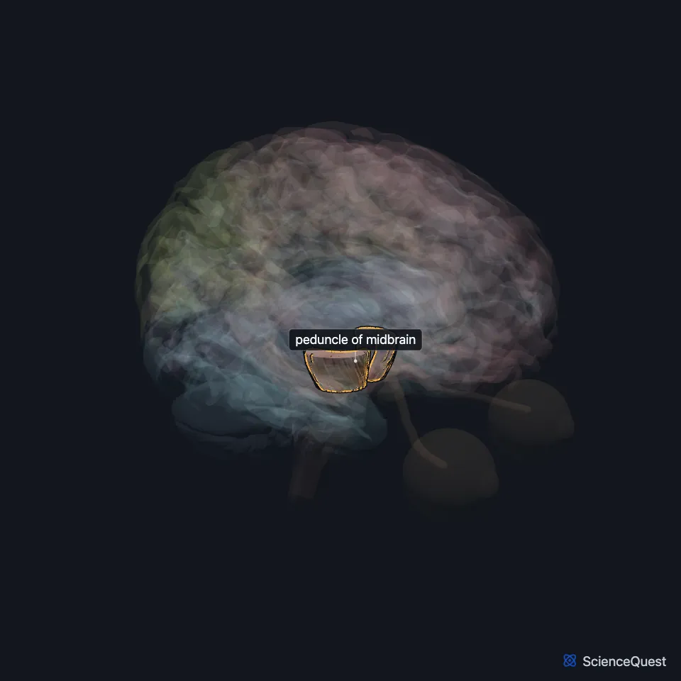
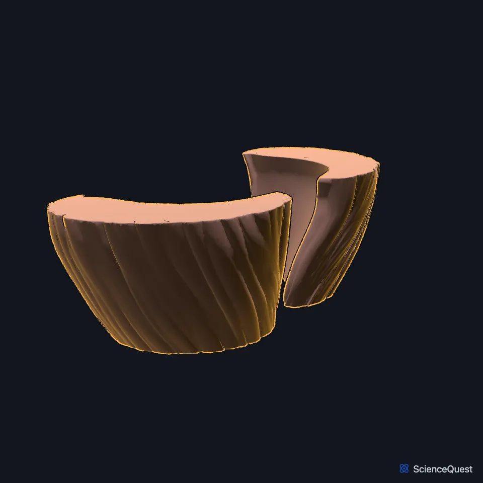
Where are the cerebral peduncles?
They rise from the upper surface of the pons, one on each side of the midline, and spread apart as they climb into the cerebral hemispheres. The hollow between them is the interpeduncular fossa. (Gray 1918, 4b. The Mid-brain; StatPearls, Brainstem)
What is a cerebral peduncle made of?
Each peduncle has a back part, the tegmentum, and a front part, the base, with a dark layer of gray matter, the substantia nigra, between them. Gray’s 1918 also calls the base the crusta. (Gray 1918, 4b. The Mid-brain; FMA, FMA62394 regional parts)
The base is almost all descending fibers from the cortex. Corticospinal fibers take its middle three fifths, with frontopontine fibers on the inner side and temporopontine fibers on the outer side, both ending in the nuclei of the pons. (Gray 1918, 4b. The Mid-brain; StatPearls, Internal Capsule)
What does the cerebral peduncle do?
It is the corticospinal tract’s path through the midbrain. The tract leaves the cortex, passes the internal capsule and the cerebral peduncles, then the pons and medulla, and most of its fibers cross to the other side in the lower medulla. (StatPearls, Pyramidal Tract and Mesencephalon Midbrain)
What lies around the cerebral peduncle?
The oculomotor nerve leaves from a groove on the inner face of each peduncle, the optic tract winds around its outer side, and the trochlear nerve crosses its side surface. (Gray 1918, 4b. The Mid-brain; StatPearls, Brainstem)
Its front surface is crossed by the superior cerebellar and posterior cerebral arteries. (Gray 1918, 4b. The Mid-brain)
What happens when a cerebral peduncle is damaged?
Weber syndrome, a midbrain stroke usually from a blocked branch of the posterior cerebral artery, hits the oculomotor fibers and the peduncle together. It gives a third nerve palsy on the same side and weakness of the opposite side of the body. (StatPearls, Weber Syndrome)
In uncal herniation the temporal lobe presses the peduncle against the edge of the tentorium, weakening the opposite side. When the far peduncle is pushed against the tentorium instead, the Kernohan notch, the weakness falls on the same side as the mass. (StatPearls, Uncal Herniation)
Where the sources differ
Gray’s 1918 gives crus cerebri as another name for the whole cerebral peduncle, tegmentum included. StatPearls uses crus cerebri for the front part of the midbrain that the corticospinal tract runs through, and the Foundational Model of Anatomy calls that part the base of the midbrain peduncle. (Gray 1918, 4b. The Mid-brain; StatPearls, Brainstem and Mesencephalon Midbrain; FMA, FMA62394 regional parts)
Touches in this model
Measured on the BodyParts3D meshes: surfaces within 2 mm of the peduncle of midbrain, nearest first, 12 of 26.
- Anterior limb of left internal capsule in contact
- Anterior limb of right internal capsule in contact
- Hypothalamus in contact
- Left medial geniculate body in contact
- Left optic tract in contact
- Left parahippocampal gyrus in contact
- Left thalamus in contact
- Mammillary body in contact
- Midbrain in contact
- Pons in contact
- Right medial geniculate body in contact
- Right optic tract in contact
Sources
Sources read between and
- Foundational Model of Anatomy, via the EMBL-EBI Ontology Lookup Service, Cerebral peduncle (FMA62394), preferred name and synonyms. Not printed on the page. Read .
- Gray’s Anatomy of the Human Body, 20th US edition (1918), IX. Neurology, 4b. The Mid-brain or Mesencephalon. Henry Gray. Public domain. Read .
- StatPearls, Neuroanatomy, Brainstem. Hayden Basinger; Jeffery P. Hogg. Last update . CC BY-NC-ND 4.0. Read .
- StatPearls, Neuroanatomy, Mesencephalon Midbrain. Francheska Caminero; Marco Cascella. Last update . CC BY-NC-ND 4.0. Read .
- Foundational Model of Anatomy, via the EMBL-EBI Ontology Lookup Service, Cerebral peduncle (FMA62394), regional parts. Not printed on the page. Read .
- StatPearls, Neuroanatomy, Internal Capsule. Marc Christopher Emos; Mahammed Z. Khan Suheb; Sanjeev Agarwal. Last update . CC BY-NC-ND 4.0. Read .
- StatPearls, Neuroanatomy, Pyramidal Tract. Qais AbuHasan; Sunil Munakomi. Last update . CC BY-NC-ND 4.0. Read .
- StatPearls, Weber Syndrome. Sunil Munakomi; Joe M. Das. Last update . CC BY-NC-ND 4.0. Read .
- StatPearls, Uncal Herniation. Walter A. Hall; Sunil Munakomi. Last update . CC BY-NC-ND 4.0. Read .
The values are facts and free to use; this page’s selection and presentation are © 2026 ScienceQuest.