Lateral geniculate body
The lateral geniculate body, also called the lateral geniculate nucleus, is the thalamic relay for sight. Most optic tract fibers end here, and its cells pass the signal on to the primary visual cortex. (FMA, FMA62209; Neuroscience Online, Visual Processing: Cortical Pathways; StatPearls, Nucleus Lateral Geniculate)
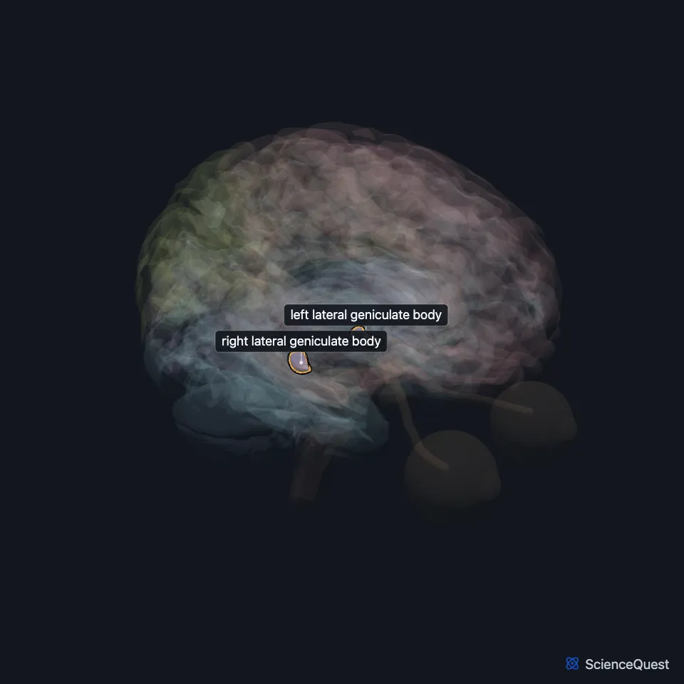
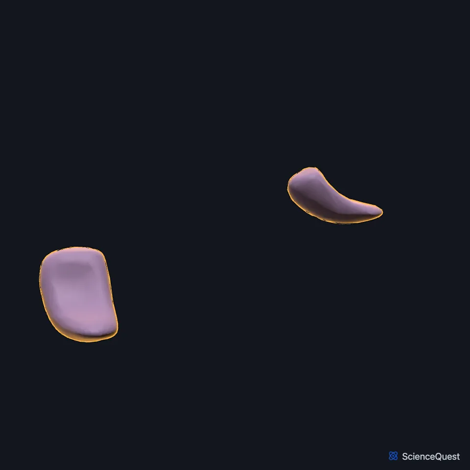
Where is the lateral geniculate body?
It sits low and at the back of the thalamus, right against the pulvinar, and is named for its place beside the medial geniculate body and for the sharp bend of its layers, from the Latin geniculum, a joint. (StatPearls, Nucleus Lateral Geniculate)
How is the lateral geniculate body layered?
It has six main layers: two deep magnocellular layers of large cells and four parvocellular layers of smaller cells above them, with thin koniocellular layers in between. (Neuroscience Online, Visual Processing: Cortical Pathways; StatPearls, Nucleus Lateral Geniculate)
Each layer takes input from one eye only. Fibers from the eye on the same side end in layers 2, 3 and 5, and those from the opposite eye in layers 1, 4 and 6. (StatPearls, Nucleus Lateral Geniculate; Neuroscience Online, Visual Processing: Cortical Pathways)
What does the lateral geniculate body do?
Its magnocellular cells take input from large-field, motion-sensitive retinal ganglion cells, and its parvocellular cells from small-field, color-sensitive ones. Far more of its input comes from higher regions, such as feedback from the visual cortex, than from the retina. (StatPearls, Nucleus Lateral Geniculate)
What connects to the lateral geniculate body?
The optic tract brings it the opposite half of the visual field from both eyes, and its output, the optic radiation, runs through the internal capsule to the visual cortex of the occipital lobe. (Neuroscience Online, Visual Processing: Cortical Pathways; StatPearls, Nucleus Lateral Geniculate and Internal Capsule)
A band of fibers, the superior brachium, links it to the superior colliculus. A distinct part between its dorsal and ventral regions, the intergeniculate leaflet, projects to the hypothalamus and pineal body, linking it to daily rhythms. (Gray 1918, 4c. The Fore-brain; StatPearls, Nucleus Lateral Geniculate)
What does damage to the lateral geniculate body cause?
A lesion here, for example from an arteriovenous malformation, causes a homonymous hemianopia or quadrantanopia in the opposite visual field. It has two blood supplies, from the posterior cerebral artery and from the anterior choroidal artery of the internal carotid. (StatPearls, Nucleus Lateral Geniculate)
Touches in this model
Measured on the BodyParts3D meshes: surfaces within 2 mm of the lateral geniculate body, nearest first, 12 of 13.
- Brachium of right superior colliculus in contact
- Left hippocampus in contact
- Left medial geniculate body in contact
- Left optic tract in contact
- Left thalamus in contact
- Right medial geniculate body in contact
- Right optic tract in contact
- Right thalamus in contact
- Brachium of left superior colliculus 0.2 mm away
- Peduncle of midbrain 0.5 mm away
- Brachium of right inferior colliculus 1.1 mm away
- Right hippocampus 1.1 mm away
Sources
Sources read
- Foundational Model of Anatomy, via the EMBL-EBI Ontology Lookup Service, Lateral geniculate body (FMA62209), preferred name and synonyms. Not printed on the page. Read .
- Neuroscience Online, McGovern Medical School, Section 2, Chapter 15, Visual Processing: Cortical Pathways. Valentin Dragoi, Ph.D. (content provided by Chieyeko Tsuchitani, Ph.D.). Reviewed . Open access; no reuse licence stated. Read .
- StatPearls, Neuroanatomy, Nucleus Lateral Geniculate. Benjamin P. Covington; Yasir Al Khalili. Last update . CC BY-NC-ND 4.0. Read .
- StatPearls, Neuroanatomy, Internal Capsule. Marc Christopher Emos; Mahammed Z. Khan Suheb; Sanjeev Agarwal. Last update . CC BY-NC-ND 4.0. Read .
- Gray’s Anatomy of the Human Body, 20th US edition (1918), IX. Neurology, 4c. The Fore-brain or Prosencephalon. Henry Gray. Public domain. Read .
The values are facts and free to use; this page’s selection and presentation are © 2026 ScienceQuest.