Posterior commissure
The posterior commissure is a rounded band of white matter that crosses the midline above the upper end of the cerebral aqueduct. Gray’s 1918 counts it, with the habenular trigone (trigonum habenulae) and the pineal body, as part of the epithalamus. (Gray 1918, 4c. The Fore-brain; FMA, FMA74868)
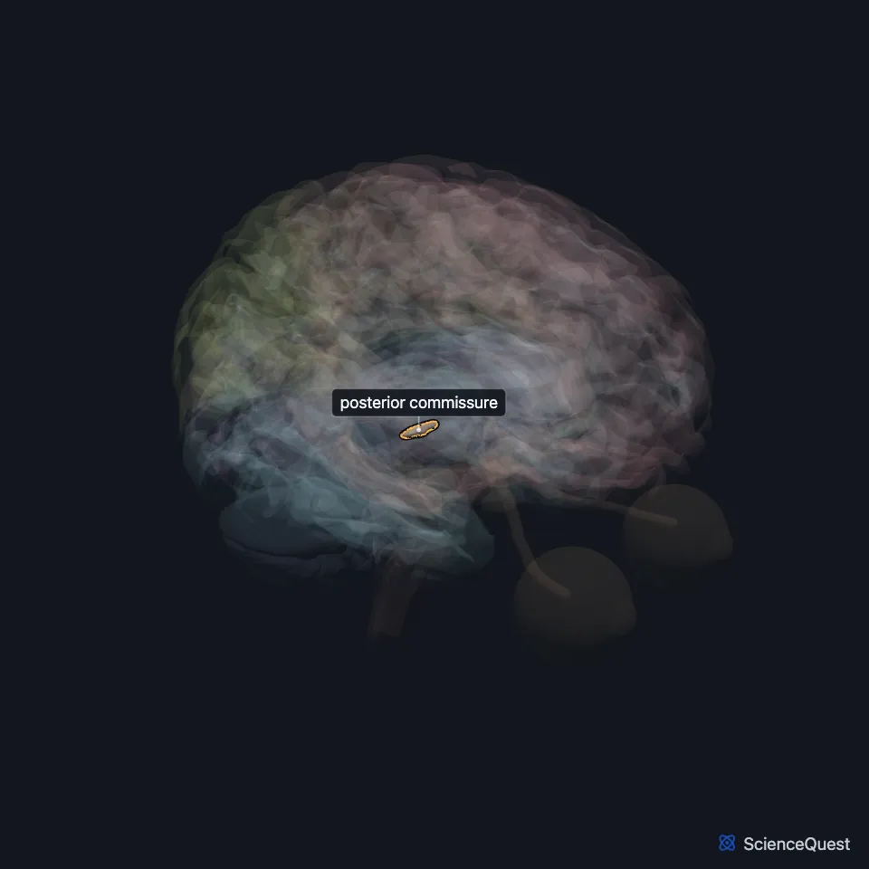
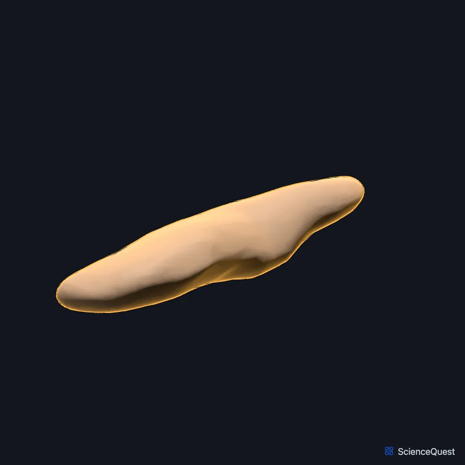
Where is the posterior commissure?
It forms part of the back wall of the third ventricle, which from top to bottom is the pineal body, the posterior commissure and the opening of the cerebral aqueduct. The pineal region lies above and behind it. (StatPearls, Ventricular System and Pineal Region Tumors; Gray 1918, 4c. The Fore-brain)
What lies next to the posterior commissure?
The first part of the cerebral aqueduct, the pars anterior, lies just under it. The superior colliculi and the rest of the corpora quadrigemina sit below and behind it. (StatPearls, Cerebral Aqueduct (Sylvian); Gray 1918, 4b. The Mid-brain)
The stalk of the pineal body splits into two sheets around the pineal recess of the third ventricle. The lower sheet is continuous with the posterior commissure, and the upper one with the habenular commissure of the habenula. (Gray 1918, 4c. The Fore-brain)
Where do the posterior commissure’s fibers come from?
Gray’s 1918 traces most of its fibers to a nucleus of the posterior commissure in the gray matter at the top of the aqueduct, and some to the thalamus and superior colliculus. It adds that their connections were not yet settled. (Gray 1918, 4c. The Fore-brain)
What does the posterior commissure do?
In the pupillary light reflex, fibers from the pretectal nucleus cross in it to reach the Edinger-Westphal nucleus of the other side. This crossing is one reason light in one eye narrows both pupils. (StatPearls, Parinaud Syndrome and Pupillary Light Reflexes and Pathway)
Fibers for upward gaze are thought to cross here too, which may make them more vulnerable to pressure from a mass. (StatPearls, Parinaud Syndrome)
What happens when the posterior commissure is compressed?
Parinaud syndrome, the dorsal midbrain syndrome, joins weak upward gaze, convergence-retraction nystagmus and pupils that react poorly to light but still narrow for near vision. Lid retraction, the Collier sign, is put down to damage of fibers at the posterior commissure. (StatPearls, Parinaud Syndrome)
Tumors of the pineal region, above it, often present with Parinaud syndrome and hydrocephalus. (StatPearls, Pineal Region Tumors)
Touches in this model
Measured on the BodyParts3D meshes: surfaces within 2 mm of the posterior commissure, nearest first.
- Left thalamus in contact
- Midbrain in contact
- Right thalamus in contact
- Third ventricle in contact
- Habenula 0.3 mm away
- Cerebral aqueduct 0.5 mm away
- Left superior colliculus 0.5 mm away
- Right superior colliculus 0.5 mm away
- Pineal body 1.0 mm away
- Brachium of left superior colliculus 1.2 mm away
- Brachium of right superior colliculus 1.2 mm away
Sources
Sources read between and
- Gray’s Anatomy of the Human Body, 20th US edition (1918), IX. Neurology, 4c. The Fore-brain or Prosencephalon. Henry Gray. Public domain. Read .
- Foundational Model of Anatomy, via the EMBL-EBI Ontology Lookup Service, Habenular trigone (FMA74868), definition and Latin names. Not printed on the page. Read .
- StatPearls, Neuroanatomy, Ventricular System. Saraswati Satyanarayan Shenoy; Forshing Lui. Last update . CC BY-NC-ND 4.0. Read .
- StatPearls, Pineal Region Tumors. Walter A. Hall; Torin Karsonovich. Last update . CC BY-NC-ND 4.0. Read .
- StatPearls, Neuroanatomy, Cerebral Aqueduct (Sylvian). Jessica M. Rubino; Jeffery P. Hogg. Last update . CC BY-NC-ND 4.0. Read .
- Gray’s Anatomy of the Human Body, 20th US edition (1918), IX. Neurology, 4b. The Mid-brain or Mesencephalon. Henry Gray. Public domain. Read .
- StatPearls, Parinaud Syndrome. Kaberi B. Feroze; Bhupendra C. Patel. Last update . CC BY-NC-ND 4.0. Read .
- StatPearls, Neuroanatomy, Pupillary Light Reflexes and Pathway. Umut Akova; Hannah Yoo; Marjorie V. Launico. Last update . CC BY-NC-ND 4.0. Read .
The values are facts and free to use; this page’s selection and presentation are © 2026 ScienceQuest.