Habenula
The habenula is a small part of the epithalamus that lies near the pineal body and the third ventricle. Its name is Latin for little rein. (FMA, FMA62032; Velasquez et al. 2014, Front Hum Neurosci)
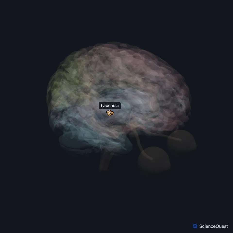
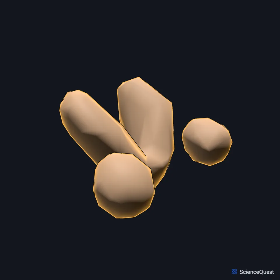
Where is the habenula?
It lies above the back end of the thalamus, close to the midline, just in front of the superior colliculus. The habenular nuclei sit in front of and above the pineal body. (Velasquez et al. 2014, Front Hum Neurosci; Gray 1918, 4c. The Fore-brain; StatPearls, Pineal Region Tumors)
What is the habenula made of?
It has two distinct nuclei, the medial habenula and the lateral habenula, and a habenular commissure that crosses the midline to the habenula of the other side. (FMA, FMA62032; Velasquez et al. 2014, Front Hum Neurosci; Gray 1918, 4c. The Fore-brain)
The pineal body sits just behind the habenular commissure. Gray’s 1918 counts three parts in the epithalamus: the habenular trigone (trigonum habenulae), the pineal body and the posterior commissure. The habenular trigone is a small triangle that holds the habenular nuclei. (StatPearls, Physiology, Pineal Gland; Gray 1918, 4c. The Fore-brain; FMA, FMA74868)
What does the habenula do?
The lateral habenula fires more when an expected reward fails to arrive, and it holds back the dopamine neurons of the midbrain and the serotonin neurons of the raphe nuclei. It takes part in how the brain handles reward and disappointment. (Browne et al. 2018, Front Synaptic Neurosci; Velasquez et al. 2014, Front Hum Neurosci)
What connects to the habenula?
Its main input arrives through the stria medullaris, a fiber bundle along the roof of the third ventricle, from the septum, hippocampus, hypothalamus and basal ganglia, including the globus pallidus. (Velasquez et al. 2014, Front Hum Neurosci)
Its output runs down the fasciculus retroflexus to the midbrain. Fibers from the medial habenula end in the interpeduncular nucleus, and the lateral habenula projects to a small inhibitory nucleus, the rostromedial tegmental nucleus, which in turn projects to the midbrain’s dopamine neurons. (Velasquez et al. 2014, Front Hum Neurosci; Gray 1918, 4b. The Mid-brain)
Why is the habenula studied in depression and addiction?
Habenular activity is tied to depression in people and to depressive-like behavior in rodents, and one review proposes that habenular overactivity contributes to major depression. In rodents, the fasciculus retroflexus degenerates after chronic exposure to cocaine, amphetamines and nicotine, and deep brain stimulation of the habenula cut cocaine seeking. (Browne et al. 2018, Front Synaptic Neurosci; Velasquez et al. 2014, Front Hum Neurosci)
Touches in this model
Measured on the BodyParts3D meshes: surfaces within 2 mm of the habenula, nearest first.
- Left stria medullaris of thalamus in contact
- Pineal body in contact
- Right stria medullaris of thalamus in contact
- Third ventricle in contact
- Posterior commissure 0.3 mm away
- Midbrain 1.0 mm away
- Choroid plexus of left cerebral hemisphere 1.4 mm away
- Choroid plexus of right cerebral hemisphere 1.4 mm away
- Left thalamus 1.4 mm away
- Right thalamus 1.5 mm away
- Right superior colliculus 1.9 mm away
Sources
Sources read between and
- Foundational Model of Anatomy, via the EMBL-EBI Ontology Lookup Service, Habenula (FMA62032), definition and synonyms. Not printed on the page. Read . Rechecked .
- Frontiers in Human Neuroscience, The role of the habenula in drug addiction, Front Hum Neurosci 8, article 174 (full text, PMC3975120). Velasquez KM, Molfese DL, Salas R. CC BY. Read .
- Gray’s Anatomy of the Human Body, 20th US edition (1918), IX. Neurology, 4c. The Fore-brain or Prosencephalon. Henry Gray. Public domain. Read .
- StatPearls, Pineal Region Tumors. Walter A. Hall; Torin Karsonovich. Last update . CC BY-NC-ND 4.0. Read .
- StatPearls, Physiology, Pineal Gland. Sadia Ilahi; Nitya Beriwal; Tahir B. Ilahi. Last update . CC BY-NC-ND 4.0. Read .
- Foundational Model of Anatomy, via the EMBL-EBI Ontology Lookup Service, Habenular trigone (FMA74868), definition and Latin names. Not printed on the page. Read .
- Frontiers in Synaptic Neuroscience, Dysregulation of the Lateral Habenula in Major Depressive Disorder, Front Synaptic Neurosci 10, article 46 (full text, PMC6292991). Browne CA, Hammack R, Lucki I. CC BY. Read . Rechecked .
- Gray’s Anatomy of the Human Body, 20th US edition (1918), IX. Neurology, 4b. The Mid-brain or Mesencephalon. Henry Gray. Public domain. Read .
The values are facts and free to use; this page’s selection and presentation are © 2026 ScienceQuest.