Septum pellucidum
The septum pellucidum is a thin, triangular double membrane that separates the front horns of the right and left lateral ventricles. Its name is Latin for translucent wall. (StatPearls, Cavum Septi Pellucidi and Ventricular System)
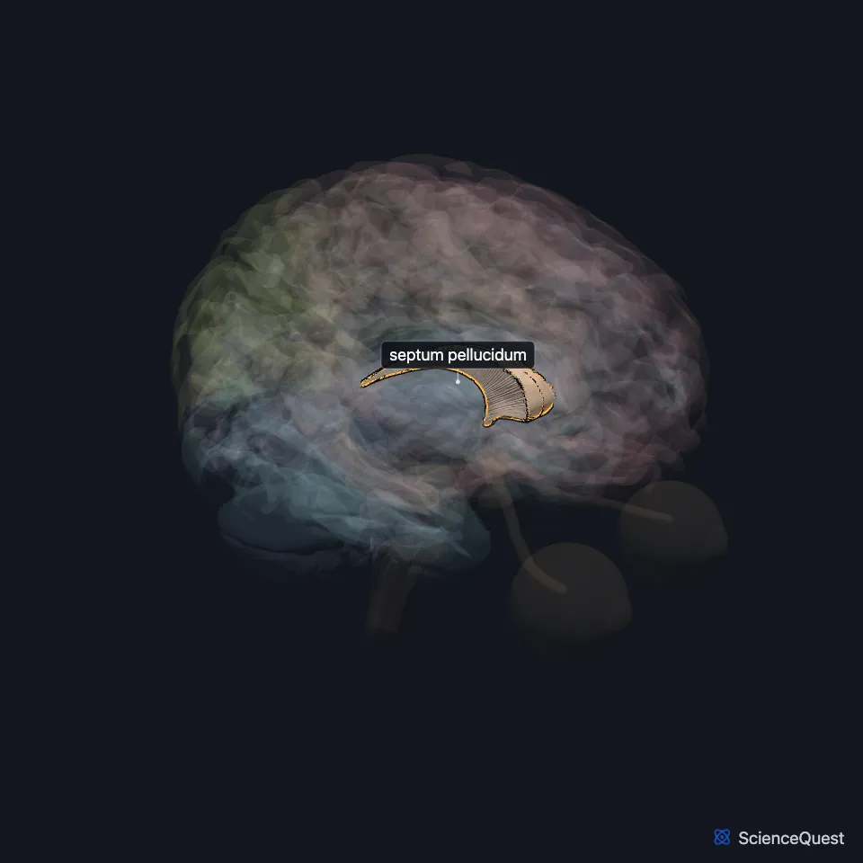
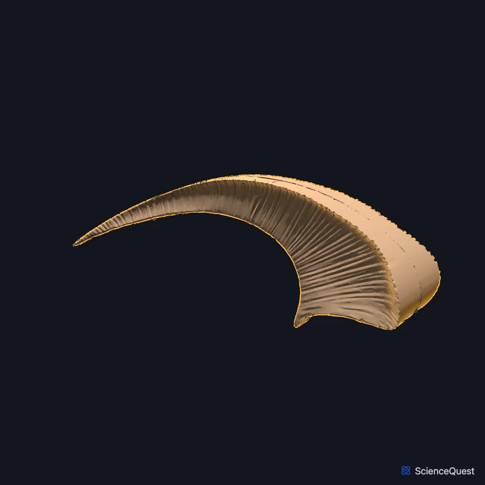
Where is the septum pellucidum?
It hangs in the midline from the underside of the corpus callosum down to the fornix. It is broad in front and narrow behind. (Gray 1918, 4c. The Fore-brain; StatPearls, Cavum Septi Pellucidi)
It forms the inner wall of the anterior horn of each lateral ventricle and most of the inner wall of its central part. (StatPearls, Ventricular System)
What is the septum pellucidum made of?
It is 1.5 to 3.0 mm thick in all, and made of two thin leaves with a narrow slit between them over most of their extent. The ependyma of the lateral ventricle covers the outer face of each leaf. (Gray 1918, 4c. The Fore-brain; StatPearls, Cavum Septi Pellucidi)
What is the septum pellucidum part of?
It walls the two lateral ventricles off from each other. StatPearls lists it among the forebrain parts of the limbic system, and Gray’s 1918 puts it in the rhinencephalon, an older grouping. (StatPearls, Ventricular System and Limbic System; Gray 1918, 4c. The Fore-brain)
What is the cavum septi pellucidi?
The gap between the two leaves is the cavum septi pellucidi. It holds fluid like cerebrospinal fluid but is not part of the ventricular system: it has no ependyma or choroid plexus and does not connect with the ventricles. (StatPearls, Cavum Septi Pellucidi; Gray 1918, 4c. The Fore-brain)
How common is an open cavum septi pellucidi?
The two leaves begin fusing at about 6 months of gestation, from back to front. A cavum is found in all premature infants and 85% of full-term newborns, usually closes 3 to 6 months after birth, and is reported in 4% to 74% of adults depending on how it is looked for. (StatPearls, Cavum Septi Pellucidi)
On its own an open cavum is usually a harmless normal variant. A missing cavum on a fetal scan is different, and prompts checks for septo-optic dysplasia and other brain malformations. (StatPearls, Cavum Septi Pellucidi)
Where the sources differ
Gray’s 1918 gives the fifth ventricle as another name for the cavity of the septum pellucidum. StatPearls calls that name incorrect, since the cavity is not part of the ventricular system. (Gray 1918, 4c. The Fore-brain; StatPearls, Cavum Septi Pellucidi)
Touches in this model
Measured on the BodyParts3D meshes: surfaces within 2 mm of the septum pellucidum, nearest first, 12 of 15.
- Anterior commissure in contact
- Corpus callosum in contact
- Left lateral ventricle in contact
- Right lateral ventricle in contact
- Third ventricle in contact
- Hypothalamus 0.1 mm away
- Left fornix of forebrain 0.3 mm away
- Right fornix of forebrain 0.3 mm away
- Choroid plexus of left cerebral hemisphere 0.4 mm away
- Choroid plexus of right cerebral hemisphere 0.4 mm away
- Lamina terminalis 0.5 mm away
- Commissure of fornix of forebrain 0.6 mm away
Sources
Sources read
- StatPearls, Cavum Septi Pellucidi. Joe M. Das; Erin Gillespie. Last update . CC BY-NC-ND 4.0. Read .
- StatPearls, Neuroanatomy, Ventricular System. Saraswati Satyanarayan Shenoy; Forshing Lui. Last update . CC BY-NC-ND 4.0. Read .
- Gray’s Anatomy of the Human Body, 20th US edition (1918), IX. Neurology, 4c. The Fore-brain or Prosencephalon. Henry Gray. Public domain. Read .
- StatPearls, Neuroanatomy, Limbic System. Tyler J. Torrico; Sara Abdijadid. Last update . CC BY-NC-ND 4.0. Read .
The values are facts and free to use; this page’s selection and presentation are © 2026 ScienceQuest.