Calcaneal tendon
The calcaneal tendon, better known as the Achilles tendon, joins the gastrocnemius and soleus to the heel bone. It is the thickest tendon in the human body. (StatPearls, Achilles Tendon)
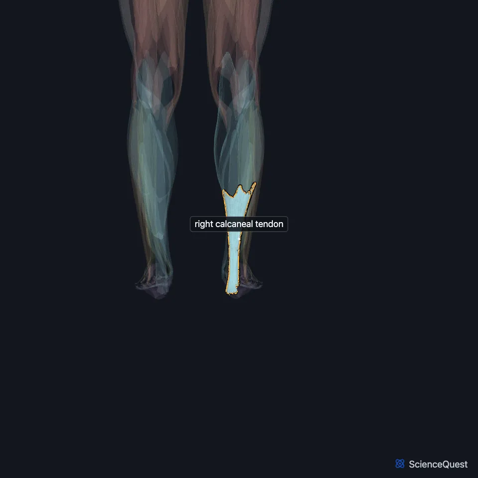
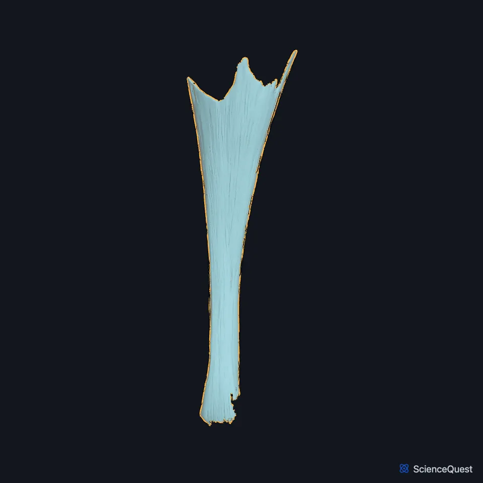
Open it in the Lower Limb Muscles Explorer See it alone in 3D
Where is the calcaneal tendon?
It lies at the back of the lower leg. Gray’s 1918 gives it a length of about 15 cm, beginning near the middle of the leg, with muscle fibers joining its front almost to its lower end. (StatPearls, Achilles Tendon; Gray’s Anatomy (1918), Posterior Crural Muscles, superficial group)
It inserts on the middle part of the back surface of the calcaneus, with a bursa between the tendon and the upper part of that surface. (StatPearls, Achilles Tendon; Gray’s Anatomy (1918), Posterior Crural Muscles, superficial group)
How is the calcaneal tendon built?
Type I collagen, which gives the tendon its strength, makes up most of it, along with elastin. It has no true synovial sheath; a paratenon of loose connective tissue surrounds it and stretches 2 to 3 cm with movement so the tendon can glide. (StatPearls, Achilles Tendon)
It spreads out a little at its lower end, so its narrowest part sits about 4 cm above the insertion. (Gray’s Anatomy (1918), Posterior Crural Muscles, superficial group)
Two bursae let it glide without friction. A subcutaneous bursa lies between the tendon and the superficial fascia over it, and a deeper retrocalcaneal bursa between the tendon and the top of the calcaneal tuberosity. (StatPearls, Leg Posterior Compartment)
What does the calcaneal tendon do?
It carries the pull of the gastrocnemius and soleus to the heel and so plantarflexes the foot for walking, running and jumping. In those movements it bears the highest loads in the body, up to ten times body weight. (StatPearls, Achilles Tendon)
A tap on the tendon makes the foot plantarflex. Doctors use this reflex to test the S1 and S2 nerve roots. (StatPearls, Achilles Tendon)
Why does the calcaneal tendon rupture?
It ruptures more often than any other tendon. A sudden force, often an eccentric load, tears it, and after a rupture, squeezing the calf (the Thompson test) gives weak or absent plantarflexion. (StatPearls, Achilles Tendon)
Its blood supply falls and it stiffens with age. The typical age at rupture is 40 to 50 years. (StatPearls, Achilles Tendon)
What supplies the calcaneal tendon, and what lies beside it?
The posterior tibial artery supplies its upper and lower sections and the fibular artery its middle section. A zone with few vessels, about 2 to 6 cm above the insertion, matches the site of many injuries. (StatPearls, Achilles Tendon)
The sural nerve gives it most of its sensory supply. The small saphenous vein runs along its lateral side, superficial to it. (StatPearls, Achilles Tendon; Gray’s Anatomy (1918), Posterior Crural Muscles, superficial group)
The plantaris inserts beside it on the calcaneus. The nearby flexor hallucis longus tendon is the one surgeons transfer most when the calcaneal tendon cannot be repaired. (StatPearls, Leg Posterior Compartment and Calf Flexor Hallucis Longus Muscle)
Where the sources differ
Sources disagree on whether it is the strongest tendon: Gray’s 1918 and the StatPearls Achilles tendon article call it the strongest tendon in the body, while the StatPearls posterior compartment article calls it among the strongest. (Gray’s Anatomy (1918), Posterior Crural Muscles, superficial group; StatPearls, Achilles Tendon and Leg Posterior Compartment)
| Feature | Detail and source |
|---|---|
| Formed by | Gastrocnemius and soleus (StatPearls, Achilles Tendon) |
| Inserts on | Middle part of the back surface of the calcaneus (StatPearls, Achilles Tendon) |
| Length | About 15 cm (Gray’s 1918) (Gray’s Anatomy (1918), Posterior Crural Muscles, superficial group) |
| Sensory nerve | Sural nerve, with small tibial nerve branches (StatPearls, Achilles Tendon) |
Touches in this model
Measured on the BodyParts3D meshes: surfaces within 2 mm of the calcaneal tendon, nearest first.
- Lateral head of right gastrocnemius in contact
- Medial head of right gastrocnemius in contact
- Right plantaris in contact
- Right soleus in contact
- Right calcaneus in contact
- Skin in contact
Sources
Sources read
- StatPearls, Anatomy, Bony Pelvis and Lower Limb: Achilles Tendon. Michael Wong; Achraf H. Jardaly; John Kiel. Last update . CC BY-NC-ND 4.0. Read .
- Anatomy of the Human Body (Gray’s Anatomy), 20th US edition, 1918, Myology, The Muscles and Fasciae of the Leg, 2. The Posterior Crural Muscles, the superficial group, pages 482 to 483; Internet Archive scan of the 1918 Lea and Febiger printing, terminology of 1918. Henry Gray; edited by Warren H. Lewis. Public domain. Read .
- StatPearls, Anatomy, Bony Pelvis and Lower Limb: Leg Posterior Compartment. Rami Abuqubo; Steven B. Graefe; James La Rose. Last update . CC BY-NC-ND 4.0. Read .
- StatPearls, Anatomy, Bony Pelvis and Lower Limb: Calf Flexor Hallucis Longus Muscle. Christopher J. Murdock; Akul Munjal; Kofi Agyeman. Last update . CC BY-NC-ND 4.0. Read .
The values are facts and free to use; this page’s selection and presentation are © 2026 ScienceQuest.