Plantaris
The plantaris is a small muscle of the superficial posterior compartment of the leg, between the gastrocnemius and the soleus. It has a long tendinous part that can be mistaken for a nerve. (StatPearls, Leg Posterior Compartment and Calf)
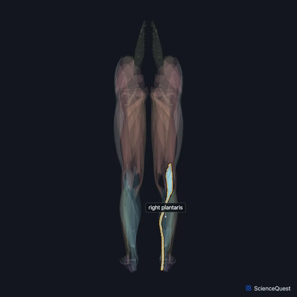
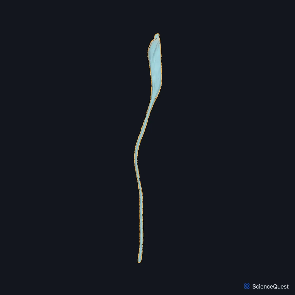
Open it in the Lower Limb Muscles Explorer See it alone in 3D
Where is the plantaris?
StatPearls places its origin on the lateral supracondylar line of the femur. Gray’s 1918 words the origin as the lower part of the lateral prolongation of the linea aspera, and adds the oblique popliteal ligament of the knee. (StatPearls, Calf; Gray’s Anatomy (1918), Posterior Crural Muscles, superficial group)
Its tendon runs between the gastrocnemius and soleus and inserts next to the calcaneal tendon on the tuberosity of the calcaneus. (StatPearls, Calf and Leg Posterior Compartment)
With the lateral head of the gastrocnemius it forms the lower lateral border of the popliteal fossa. (StatPearls, Popliteal Region)
How is the plantaris built?
Gray’s 1918 measures its spindle-shaped belly at 7 to 10 cm. StatPearls calls its tendon the longest in the body. (Gray’s Anatomy (1918), Posterior Crural Muscles, superficial group; StatPearls, Popliteal Region)
What does the plantaris do?
It helps plantarflex the ankle, far less than the gastrocnemius and soleus do. (StatPearls, Leg Posterior Compartment)
Gray’s 1918 calls it the rudiment of a large muscle that, in some other animals, continues over the heel into the plantar aponeurosis. In humans it serves as an accessory to the gastrocnemius. (Gray’s Anatomy (1918), Posterior Crural Muscles, superficial group)
Surgeons often take it as graft material. In most calcaneal tendon ruptures its muscle and tendon stay intact. (StatPearls, Leg Posterior Compartment and Gastrocnemius Muscle)
What supplies the plantaris?
The tibial nerve gives it branches in the popliteal fossa, as it does the gastrocnemius, soleus and popliteus. (StatPearls, Calf)
Gray’s 1918 traces its supply through the tibial nerve to the fourth and fifth lumbar and first sacral nerves. (Gray’s Anatomy (1918), Posterior Crural Muscles, superficial group)
The tibial nerve and the popliteal artery both pass deep to it and the gastrocnemius. (StatPearls, Tibial Nerve)
Is the plantaris always present?
About 10% of people lack it. Gray’s 1918 adds that it is sometimes double, and that its tendon may end in the laciniate ligament or the fascia of the leg. (StatPearls, Gastrocnemius Muscle; Gray’s Anatomy (1918), Posterior Crural Muscles, superficial group)
It varies in origin, course and insertion, and fetal studies describe at least 8 different insertion forms. (StatPearls, Calf)
Where the sources differ
Sources disagree on how its tendon meets the calcaneal tendon. The StatPearls calf article has it blend with that tendon and Gray’s 1918 runs it along its medial border, while the StatPearls gastrocnemius article places it lateral to the calcaneal tendon and says it seldom fuses with it. (StatPearls, Calf and Gastrocnemius Muscle; Gray’s Anatomy (1918), Posterior Crural Muscles, superficial group)
| Feature | Detail and source |
|---|---|
| Origin | Lateral supracondylar line of the femur (StatPearls, Leg Posterior Compartment) |
| Insertion | Tuberosity of the calcaneus, next to the calcaneal tendon (StatPearls, Leg Posterior Compartment) |
| Action | Weak plantarflexion of the ankle (StatPearls, Leg Posterior Compartment) |
| Nerve | Tibial nerve (StatPearls, Calf) |
Touches in this model
Measured on the BodyParts3D meshes: surfaces within 2 mm of the plantaris, nearest first.
- Lateral head of right gastrocnemius in contact
- Medial head of right gastrocnemius in contact
- Right calcaneal tendon in contact
- Right popliteus in contact
- Right soleus in contact
- Short head of right biceps femoris in contact
- Right calcaneus in contact
- Right femur in contact
- Skin in contact
- Long head of right biceps femoris 0.5 mm away
Sources
Sources read
- StatPearls, Anatomy, Bony Pelvis and Lower Limb: Leg Posterior Compartment. Rami Abuqubo; Steven B. Graefe; James La Rose. Last update . CC BY-NC-ND 4.0. Read .
- StatPearls, Anatomy, Bony Pelvis and Lower Limb: Calf. Justin T. Binstead; Matthew A. Varacallo. Last update . CC BY-NC-ND 4.0. Read .
- Anatomy of the Human Body (Gray’s Anatomy), 20th US edition, 1918, Myology, The Muscles and Fasciae of the Leg, 2. The Posterior Crural Muscles, the superficial group, pages 482 to 483; Internet Archive scan of the 1918 Lea and Febiger printing, terminology of 1918. Henry Gray; edited by Warren H. Lewis. Public domain. Read .
- StatPearls, Anatomy, Bony Pelvis and Lower Limb: Popliteal Region. Scott Hyland; Margaret A. Sinkler; Matthew A. Varacallo. Last update . CC BY-NC-ND 4.0. Read .
- StatPearls, Anatomy, Bony Pelvis and Lower Limb: Gastrocnemius Muscle. Bruno Bordoni; Matthew A. Varacallo. Last update . CC BY-NC-ND 4.0. Read .
- StatPearls, Anatomy, Bony Pelvis and Lower Limb: Tibial Nerve. Sohil S. Desai; James La Rose. Last update . CC BY-NC-ND 4.0. Read .
The values are facts and free to use; this page’s selection and presentation are © 2026 ScienceQuest.