Popliteus
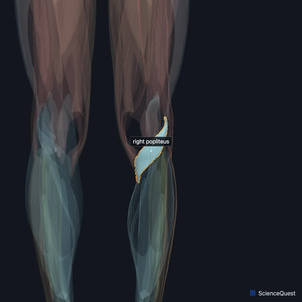
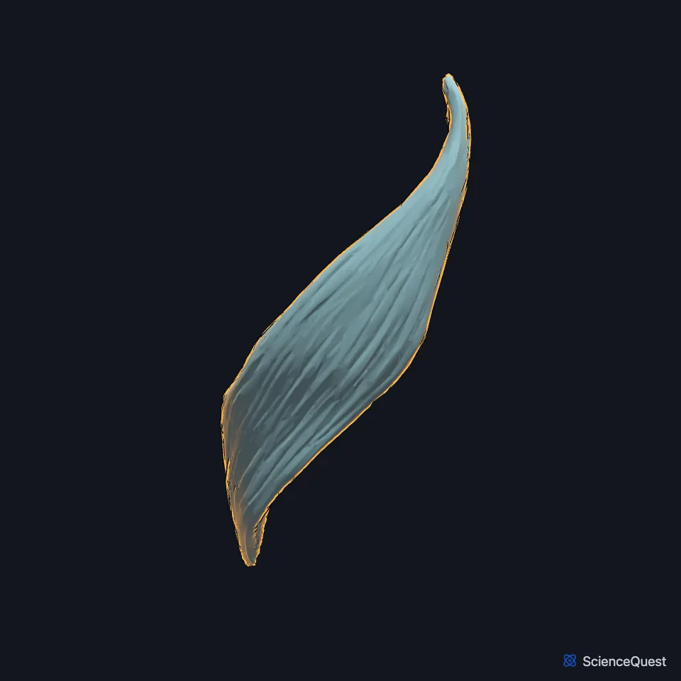
The popliteus is a thin, flat, triangular muscle behind the knee that forms the lower part of the floor of the popliteal fossa. It unlocks the knee at full extension by rotating the tibia medially, and so starts flexion. (Gray’s Anatomy (1918), Posterior Crural Muscles, deep group; StatPearls, Leg Posterior Compartment and Calf)
Open it in the Lower Limb Muscles Explorer See it alone in 3D
Where is the popliteus?
It is the highest muscle of the deep posterior compartment of the leg, which it shares with the tibialis posterior, flexor digitorum longus and flexor hallucis longus. (StatPearls, Leg Posterior Compartment and Popliteus Muscle)
It arises from the lateral condyle of the femur and inserts on the back of the tibia above the soleal line, just above the origin of the soleus. (StatPearls, Popliteus Muscle and Leg Posterior Compartment)
How is the popliteus attached?
Its origin is a strong tendon about 2.5 cm long from a depression at the front of the groove on the lateral condyle of the femur, with a few fibers from the oblique popliteal ligament of the knee. (Gray’s Anatomy (1918), Posterior Crural Muscles, deep group)
StatPearls calls it a capsular but extra-articular structure that separates the lateral meniscus from the lateral collateral ligament. It also attaches to the lateral meniscus and draws it backward as the knee bends, so the femur and tibia do not trap it. (StatPearls, Popliteus Muscle)
What does the popliteus do?
It acts only at the knee. With the foot off the ground it rotates the tibia medially on the femur, and with the foot planted it rotates the femur laterally on the tibia; either way it unlocks the extended knee and starts flexion. (StatPearls, Leg Posterior Compartment)
In full extension under load, the femur rotates medially on the tibia and locks the knee with no muscular effort. The popliteus contracts to undo that lock, so some call it the key to the locked knee. (StatPearls, Popliteus Muscle)
It also checks excessive tibial rotation and forward translation of the knee. Heavy sprinting or downhill running can overcome that check. (StatPearls, Popliteus Muscle)
What supplies the popliteus?
The tibial nerve supplies it and enters at the lower lateral margin of the muscle, below the head of the fibula. (StatPearls, Popliteus Muscle)
The medial inferior genicular branch of the popliteal artery and a muscular branch of the posterior tibial artery supply it. The popliteal artery ends at its lower border and divides there. (StatPearls, Popliteus Muscle and Popliteal Region)
On its way down, the popliteal artery passes deep to the plantaris and the gastrocnemius to reach the surface of the popliteus. (StatPearls, Tibial Nerve)
Does the popliteus vary?
A small sesamoid bone, the cyamella, sometimes sits in its tendon. It can cause pain that mimics a posterolateral meniscal tear. (StatPearls, Popliteus Muscle)
Two uncommon extra muscles lie near it, the popliteus minor and the peroneotibialis. Gray’s 1918 puts the peroneotibialis at 14 per cent, running from the inner side of the fibular head to the tibia beneath the popliteus. (StatPearls, Popliteus Muscle; Gray’s Anatomy (1918), Posterior Crural Muscles, deep group)
Where the sources differ
Sources give different roots for its nerve: L5 to S1 in the StatPearls popliteus article, the fourth and fifth lumbar and first sacral nerves in Gray’s 1918. (StatPearls, Popliteus Muscle; Gray’s Anatomy (1918), Posterior Crural Muscles, deep group)
| Feature | Detail and source |
|---|---|
| Origin | Lateral condyle of the femur (StatPearls, Popliteus Muscle) |
| Insertion | Back of the tibia, above the soleal line (StatPearls, Popliteus Muscle) |
| Action | Unlocks the extended knee: rotates the tibia medially, or the femur laterally when the foot is planted (StatPearls, Leg Posterior Compartment) |
| Nerve | Tibial nerve (StatPearls, Popliteus Muscle) |
Touches in this model
Measured on the BodyParts3D meshes: surfaces within 2 mm of the popliteus, nearest first.
- Lateral head of right gastrocnemius in contact
- Medial head of right gastrocnemius in contact
- Right plantaris in contact
- Right semitendinosus in contact
- Right soleus in contact
- Short head of right biceps femoris in contact
- Right femur in contact
- Right tibia in contact
- Skin in contact
- Long head of right biceps femoris 0.4 mm away
Sources
Sources read
- Anatomy of the Human Body (Gray’s Anatomy), 20th US edition, 1918, Myology, The Muscles and Fasciae of the Leg, 2. The Posterior Crural Muscles, the deep group, pages 483 to 486; Internet Archive scan of the 1918 Lea and Febiger printing, terminology of 1918. Henry Gray; edited by Warren H. Lewis. Public domain. Read .
- StatPearls, Anatomy, Bony Pelvis and Lower Limb: Leg Posterior Compartment. Rami Abuqubo; Steven B. Graefe; James La Rose. Last update . CC BY-NC-ND 4.0. Read .
- StatPearls, Anatomy, Bony Pelvis and Lower Limb: Calf. Justin T. Binstead; Matthew A. Varacallo. Last update . CC BY-NC-ND 4.0. Read .
- StatPearls, Anatomy, Bony Pelvis and Lower Limb: Popliteus Muscle. Scott Hyland; Matthew A. Varacallo. Last update . CC BY-NC-ND 4.0. Read .
- StatPearls, Anatomy, Bony Pelvis and Lower Limb: Popliteal Region. Scott Hyland; Margaret A. Sinkler; Matthew A. Varacallo. Last update . CC BY-NC-ND 4.0. Read .
- StatPearls, Anatomy, Bony Pelvis and Lower Limb: Tibial Nerve. Sohil S. Desai; James La Rose. Last update . CC BY-NC-ND 4.0. Read .
The values are facts and free to use; this page’s selection and presentation are © 2026 ScienceQuest.