Lunate
The lunate is a crescent-shaped carpal bone. It sits in the middle of the proximal row of the wrist, between the scaphoid and the triquetral. (StatPearls, Hand Carpal Bones; Gray’s Anatomy (1918), Lunate bone, p. 224)
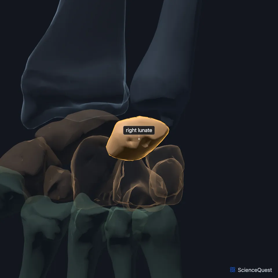
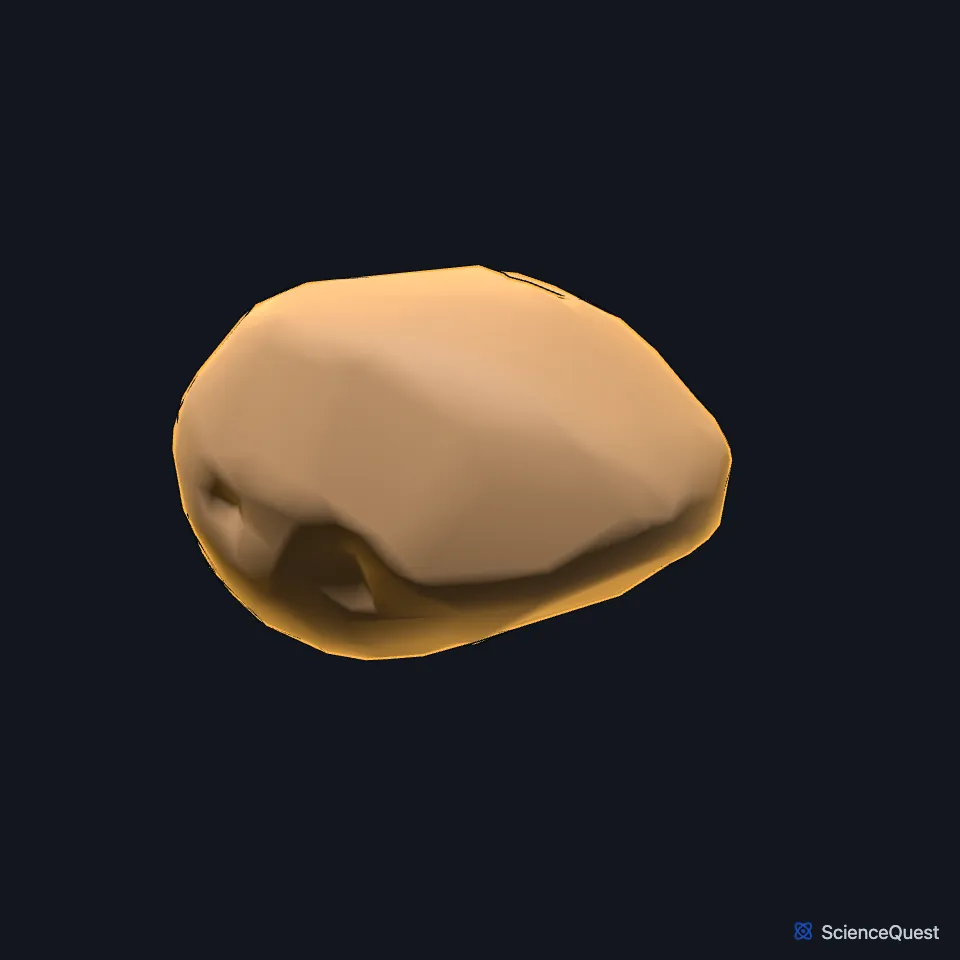
Open it in the Hand and Wrist Skeleton Explorer See it alone in 3D
Where is the lunate?
The lunate is the central bone of the proximal carpal row, with the scaphoid on its thumb side and the triquetral on its little-finger side. Its smooth, convex upper surface meets the radius. (StatPearls, Kienbock Disease and Perilunate Dislocation; Gray’s Anatomy (1918), Lunate bone, p. 224)
Which bones does the lunate meet?
Gray’s Anatomy (1918) lists five: the radius above, the capitate and hamate below, the scaphoid on the thumb side and the triquetral on the little-finger side. The head of the capitate sits in the deep hollow of its lower surface. (Gray’s Anatomy (1918), Lunate bone, p. 224)
Two ligaments inside the wrist, the scapholunate and the lunotriquetral, bind it to the scaphoid and the triquetral. (StatPearls, Perilunate Dislocation)
The lunate lies near the median nerve. A lunate pushed forward out of place can press on the nerve inside the carpal tunnel, causing tingling in the thumb, index, middle and half of the ring finger. (StatPearls, Hand Carpal Bones)
What does the lunate do?
With the scaphoid and the triquetral it forms the carpal side of the wrist joint. About 35% of the axial load across the wrist passes through the contact between the radius and the lunate, and about 10% passes to the ulna through the triangular fibrocartilage complex. (StatPearls, Hand Radiocarpal Joint and Kienbock Disease)
No tendon inserts on the scaphoid, lunate or triquetral. The three form an intercalated segment, moved only by the forces of the bones and ligaments around them. (StatPearls, Hand Radiocarpal Joint)
How does blood reach the lunate, and what is Kienböck disease?
Arteries from the arches of vessels on the back and front of the wrist enter the lunate from both surfaces, commonly two to four of them crossing the volar carpal ligaments. A smaller number of volar branches goes with a higher chance of Kienböck disease. (StatPearls, Kienbock Disease and Hand Radiocarpal Joint)
Kienböck disease is avascular necrosis of the lunate, first described by the Austrian radiologist Robert Kienböck in 1910. In some studies up to 78% of patients had an ulna short for the radius, which puts more repeated stress on the lunate. (StatPearls, Kienbock Disease)
Where the sources differ
Gray’s Anatomy (1918) counts the hamate among the lunate’s five articulations. StatPearls’ article on Kienböck disease says the lunate meets the hamate only occasionally, and sorts lunates into a more triangular type 1, with no medial articular facet, and square or rectangular types 2 and 3. (Gray’s Anatomy (1918), Lunate bone, p. 224; StatPearls, Kienbock Disease)
StatPearls’ article on the carpal bones says most lunates (80%) have two areas of blood supply joined inside the bone. Its article on Kienböck disease calls the vessels linking them inside the bone sparse. (StatPearls, Hand Carpal Bones and Kienbock Disease)
| Feature | Detail and source |
|---|---|
| Position | Proximal carpal row, centre (StatPearls, Kienbock Disease; Gray’s Anatomy (1918), Lunate bone, p. 224) |
| Articulates with | Radius, capitate, hamate, scaphoid, triquetral (Gray’s Anatomy (1918), Lunate bone, p. 224) |
| Other name | Semilunar bone, in Gray’s Anatomy (1918) (Gray’s Anatomy (1918), Lunate bone, p. 224) |
Touches in this model
Measured on the BodyParts3D meshes: surfaces within 2 mm of the lunate, nearest first.
- Right capitate in contact
- Right scaphoid in contact
- Right triquetral in contact
- Skin in contact
- Right flexor digitorum profundus 0.5 mm away
- Right hamate 0.6 mm away
- Right extensor digitorum 1.1 mm away
- Right ulna 1.2 mm away
Sources
Sources read
- StatPearls, Anatomy, Shoulder and Upper Limb, Hand Carpal Bones. Alex Tang; Matthew A. Varacallo. Last update . CC BY-NC-ND 4.0. Read .
- Gray’s Anatomy of the Human Body, 20th US edition (1918), edited by Warren H. Lewis, Osteology, The Carpus, The Lunate Bone (p. 224); 1918 names: navicular for scaphoid, triangular for triquetral. Henry Gray; Warren H. Lewis (editor). Public domain. Read .
- StatPearls, Kienbock Disease. Layla A. Nasr; Jennifer Koay. Last update . CC BY-NC-ND 4.0. Read .
- StatPearls, Perilunate Dislocation. Nicholas Frane; William Goldenberg. Last update . CC BY-NC-ND 4.0. Read .
- StatPearls, Anatomy, Shoulder and Upper Limb, Hand Radiocarpal Joint. Jackson Taylor Helms; Kenia A. Maldonado; Bracken Burns. Last update . CC BY-NC-ND 4.0. Read .
The values are facts and free to use; this page’s selection and presentation are © 2026 ScienceQuest.