Subclavian vein
The subclavian vein is the main vein draining the upper limb. It continues the axillary vein at the outer border of the first rib and ends by joining the internal jugular vein to form the brachiocephalic vein. (StatPearls, Thorax, Subclavian Veins; Gray’s Anatomy (1918), The Subclavian and Innominate Veins)
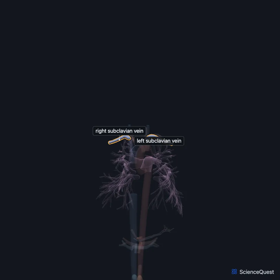
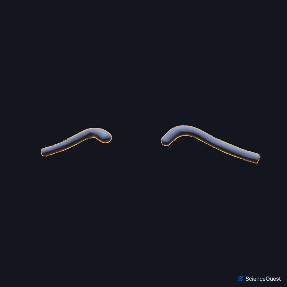
Where is the subclavian vein?
It arches behind the clavicle and joins the internal jugular vein behind the sternoclavicular joint. (StatPearls, Thorax, Subclavian Veins)
In front of it lie the clavicle and the subclavius muscle. Below, it rests in a hollow on the first rib and on the pleura. (Gray’s Anatomy (1918), The Subclavian and Innominate Veins; StatPearls, Thorax, Subclavian Veins)
What does the subclavian vein do?
Blood from the axillary vein passes through it into the brachiocephalic vein and on to the right atrium. (StatPearls, Thorax, Subclavian Veins)
Its meeting point with the internal jugular vein is the venous angle. The left angle receives the thoracic duct and the right angle the right lymphatic duct. (StatPearls, Thorax, Subclavian Veins; Gray’s Anatomy (1918), The Subclavian and Innominate Veins)
It receives the external jugular vein and sometimes the anterior jugular vein. (Gray’s Anatomy (1918), The Subclavian and Innominate Veins)
What lies next to the subclavian vein?
The subclavian artery lies behind and above it, separated from it by the anterior scalene muscle. (StatPearls, Thorax, Subclavian Veins; Gray’s Anatomy (1918), The Subclavian and Innominate Veins)
The phrenic nerve passes behind the vein, and the inferior trunk of the brachial plexus lies to its lateral side. (StatPearls, Thorax, Subclavian Veins)
Why is the subclavian vein used for central lines?
It is a common site for central venous catheters, most often by the infraclavicular route. The nearby subclavian artery, pleura and brachial plexus explain the risks, which include pneumothorax and arterial injury. (StatPearls, Thorax, Subclavian Veins)
What is Paget-Schroetter syndrome?
Paget-Schroetter syndrome is a clot in the subclavian vein caused by repeated compression. It typically affects the dominant arm of young athletes who make repeated overhead or forceful arm movements. (StatPearls, Thorax, Subclavian Veins)
Compression in the costoclavicular space is the most common site of subclavian vein compression and one of the three forms of thoracic outlet syndrome. (StatPearls, Thorax, Subclavian Veins)
Where the sources differ
The sources disagree on valves. StatPearls calls the subclavian veins valveless, while Gray’s Anatomy (1918) describes a pair of valves about 2.5 cm from the vein’s end. (StatPearls, Thorax, Subclavian Veins; Gray’s Anatomy (1918), The Subclavian and Innominate Veins)
| Feature | Detail and source |
|---|---|
| Begins | At the outer border of the first rib, as the continuation of the axillary vein (StatPearls, Thorax, Subclavian Veins; Gray’s Anatomy (1918), The Subclavian and Innominate Veins) |
| Ends | Joins the internal jugular vein to form the brachiocephalic vein (StatPearls, Thorax, Subclavian Veins) |
| Receives | External jugular vein (Gray’s Anatomy (1918), The Subclavian and Innominate Veins) |
Touches in this model
Measured on the BodyParts3D meshes: surfaces within 2 mm of the subclavian vein, nearest first, 12 of 24.
- Left brachiocephalic vein in contact
- Left internal jugular vein in contact
- Right brachiocephalic vein in contact
- Right internal jugular vein in contact
- Clavicular part of left pectoralis major in contact
- Clavicular part of right pectoralis major in contact
- External intercostal muscle in contact
- Left clavicle in contact
- Left first rib in contact
- Left serratus anterior in contact
- Left sternohyoid in contact
- Left sternothyroid in contact
Sources
Sources read
- StatPearls, Anatomy, Thorax, Subclavian Veins. Adekunle E. Omole; Vincent Sicari. Last update . CC BY-NC-ND 4.0. Read .
- Anatomy of the Human Body (Gray’s Anatomy), 20th US edition, 1918, Angiology, The Veins of the Upper Extremity and Thorax: The Subclavian Vein and The Innominate Veins, pages 664 to 666; Internet Archive scan of the 1918 Lea and Febiger printing, terminology of 1918 (innominate for brachiocephalic, internal mammary for internal thoracic). Henry Gray; edited by Warren H. Lewis. Public domain. Read .
The values are facts and free to use; this page’s selection and presentation are © 2026 ScienceQuest.