Quadratus femoris
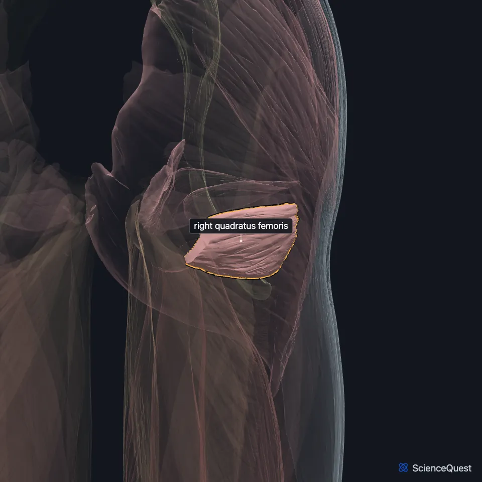
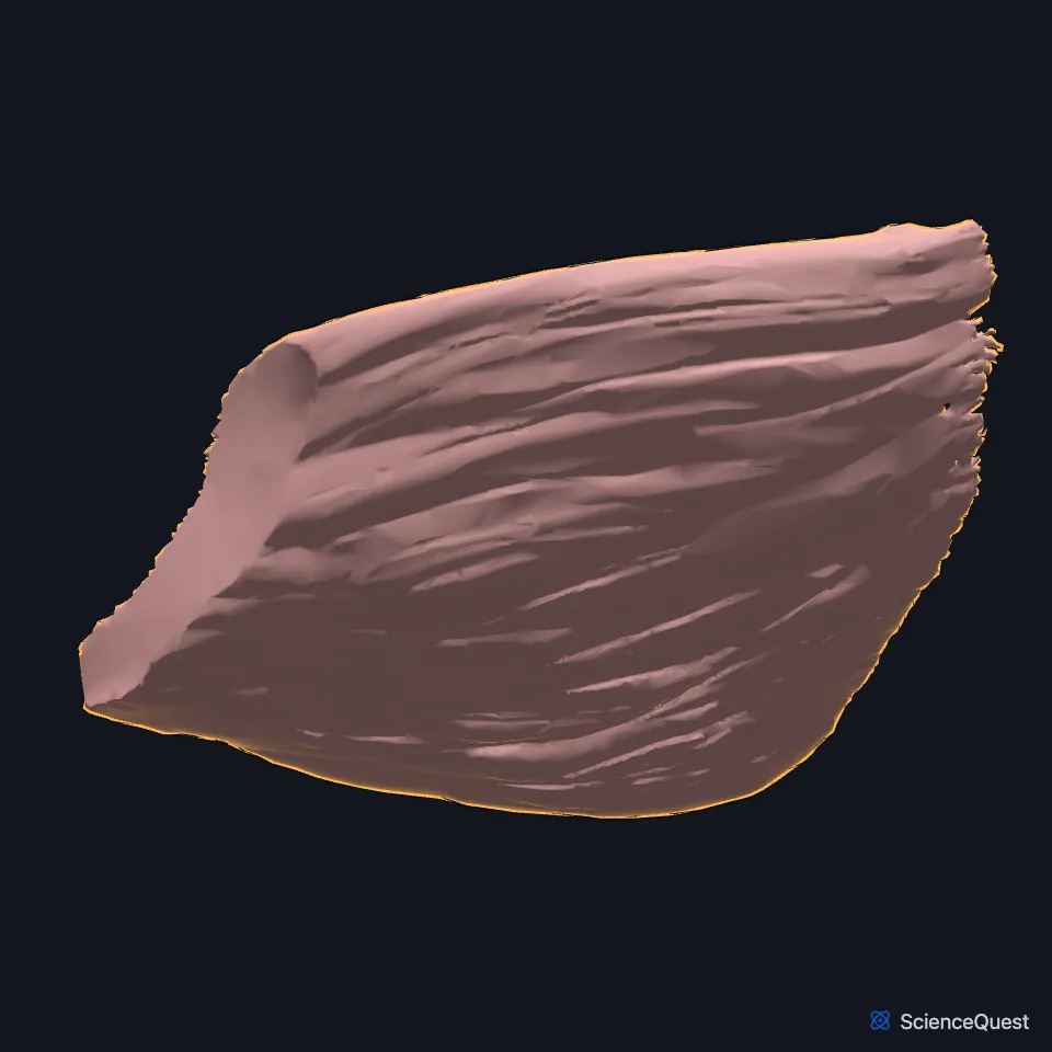
The quadratus femoris is a flat, four-sided muscle at the back of the hip, running from the ischial tuberosity to the femur. It is one of the lateral rotators of the thigh. (Gray’s Anatomy (1918), Muscles of the Gluteal Region; StatPearls, Hip Joint)
Open it in the Lower Limb Muscles Explorer See it alone in 3D
Where is the quadratus femoris?
It lies between the gemellus inferior above and the upper edge of the adductor magnus below. The end branches of the medial circumflex femoral vessels separate it from the adductor magnus. (Gray’s Anatomy (1918), Muscles of the Gluteal Region)
Where does the quadratus femoris attach?
It arises from the upper part of the outer border of the ischial tuberosity. It inserts on the upper part of what Gray’s (1918) calls the quadrate line, the line that runs straight down from the intertrochanteric crest of the femur. The StatPearls femur article names its insertion the quadrate tubercle. (Gray’s Anatomy (1918), Muscles of the Gluteal Region; StatPearls, Femur)
A bursa is often found between the front of the muscle and the lesser trochanter. (Gray’s Anatomy (1918), Muscles of the Gluteal Region)
What does the quadratus femoris do?
It rotates the thigh laterally, with the obturator muscles and the gemelli. (StatPearls, Hip Joint; Gray’s Anatomy (1918), Muscles of the Gluteal Region)
Gray’s (1918) adds that with the thigh flexed, as in sitting, these short rotators stop rotating and act as abductors. (Gray’s Anatomy (1918), Muscles of the Gluteal Region)
Which nerve and artery lie by the quadratus femoris?
Its own nerve, the nerve to the quadratus femoris, leaves the pelvis below the piriformis, runs deep to the short rotators and ends on the deep surface of the muscle. (StatPearls, Hip and Gemelli Muscles)
The deep branch of the medial circumflex femoral artery runs deep to it and shows in the gap between it and the gemellus inferior. Surgeons look for it there because the vessel is at risk in the posterior approach for hip arthroplasty. (StatPearls, Hip)
How does the quadratus femoris vary?
It is sometimes absent, and the adductor magnus may be fused with it. (Gray’s Anatomy (1918), Muscles of the Gluteal Region and Medial Femoral Muscles)
In a cadaver study StatPearls cites, where the adductor minimus was absent or small, the quadratus femoris reached lower but stayed separated from the adductor magnus by a layer of fascia. (StatPearls, Thigh Adductor Magnus Muscle)
Where the sources differ
Sources name its femoral attachment differently. The StatPearls hip article gives the intertrochanteric crest and the StatPearls femur article the quadrate tubercle; Gray’s (1918) gives the upper part of the quadrate line, its term for the line that runs down from that crest. (StatPearls, Hip and Femur; Gray’s Anatomy (1918), Muscles of the Gluteal Region)
Sources also give different roots for its nerve. The StatPearls hip article gives L4 to S1; the StatPearls femur article gives L5 and S1, which matches the last lumbar and first sacral nerves of Gray’s (1918). (StatPearls, Hip and Femur; Gray’s Anatomy (1918), Muscles of the Gluteal Region)
| Feature | Detail and source |
|---|---|
| Origin | Outer border of the ischial tuberosity, upper part (Gray’s Anatomy (1918), Muscles of the Gluteal Region; StatPearls, Hip) |
| Insertion | Upper quadrate line below the intertrochanteric crest (Gray’s 1918 term); quadrate tubercle or intertrochanteric crest (StatPearls) (Gray’s Anatomy (1918), Muscles of the Gluteal Region; StatPearls, Femur and Hip) |
| Action | Rotates the thigh laterally (StatPearls, Hip Joint) |
| Nerve | Nerve to the quadratus femoris (L4 to S1, or L5 and S1, by source) (StatPearls, Hip and Femur) |
Touches in this model
Measured on the BodyParts3D meshes: surfaces within 2 mm of the quadratus femoris, nearest first.
- Right obturator externus in contact
- Right psoas major in contact
- Right femur in contact
- Right hip bone in contact
- Skin in contact
- Right adductor magnus 1.3 mm away
Sources
Sources read
- Anatomy of the Human Body (Gray’s Anatomy), 20th US edition, 1918, Myology, The Muscles and Fasciae of the Thigh, 3. The Muscles of the Gluteal Region, pages 474 to 478; Internet Archive scan of the 1918 Lea and Febiger printing, terminology of 1918. Henry Gray; edited by Warren H. Lewis. Public domain. Read .
- StatPearls, Anatomy, Bony Pelvis and Lower Limb, Hip Joint. Maks Gold; Akul Munjal; Matthew A. Varacallo. Last update . CC BY-NC-ND 4.0. Read .
- StatPearls, Anatomy, Bony Pelvis and Lower Limb: Femur. Amy Chang; Grant Breeland; Asa C. Black; John B. Hubbard. Last update . CC BY-NC-ND 4.0. Read .
- StatPearls, Anatomy, Bony Pelvis and Lower Limb, Hip. Roland Glenister; Sandeep Sharma. Last update . CC BY-NC-ND 4.0. Read .
- StatPearls, Anatomy, Bony Pelvis and Lower Limb, Gemelli Muscles. Bradley Lezak; Dustin H. Massel. Last update . CC BY-NC-ND 4.0. Read .
- Anatomy of the Human Body (Gray’s Anatomy), 20th US edition, 1918, Myology, The Muscles and Fasciae of the Thigh, 2. The Medial Femoral Muscles, pages 471 to 474; Internet Archive scan of the 1918 Lea and Febiger printing, terminology of 1918. Henry Gray; edited by Warren H. Lewis. Public domain. Read .
- StatPearls, Anatomy, Bony Pelvis and Lower Limb: Thigh Adductor Magnus Muscle. Susan H. Jeno; Marjorie V. Launico; Gary S. Schindler. Last update . CC BY-NC-ND 4.0. Read .
The values are facts and free to use; this page’s selection and presentation are © 2026 ScienceQuest.