Obturator externus
The obturator externus is a flat, triangular muscle on the outer surface of the front wall of the pelvis. Unlike the other short lateral rotators of the hip, it is supplied from the lumbar plexus. (Gray’s Anatomy (1918), Muscles of the Gluteal Region; StatPearls, Obturator Muscles)
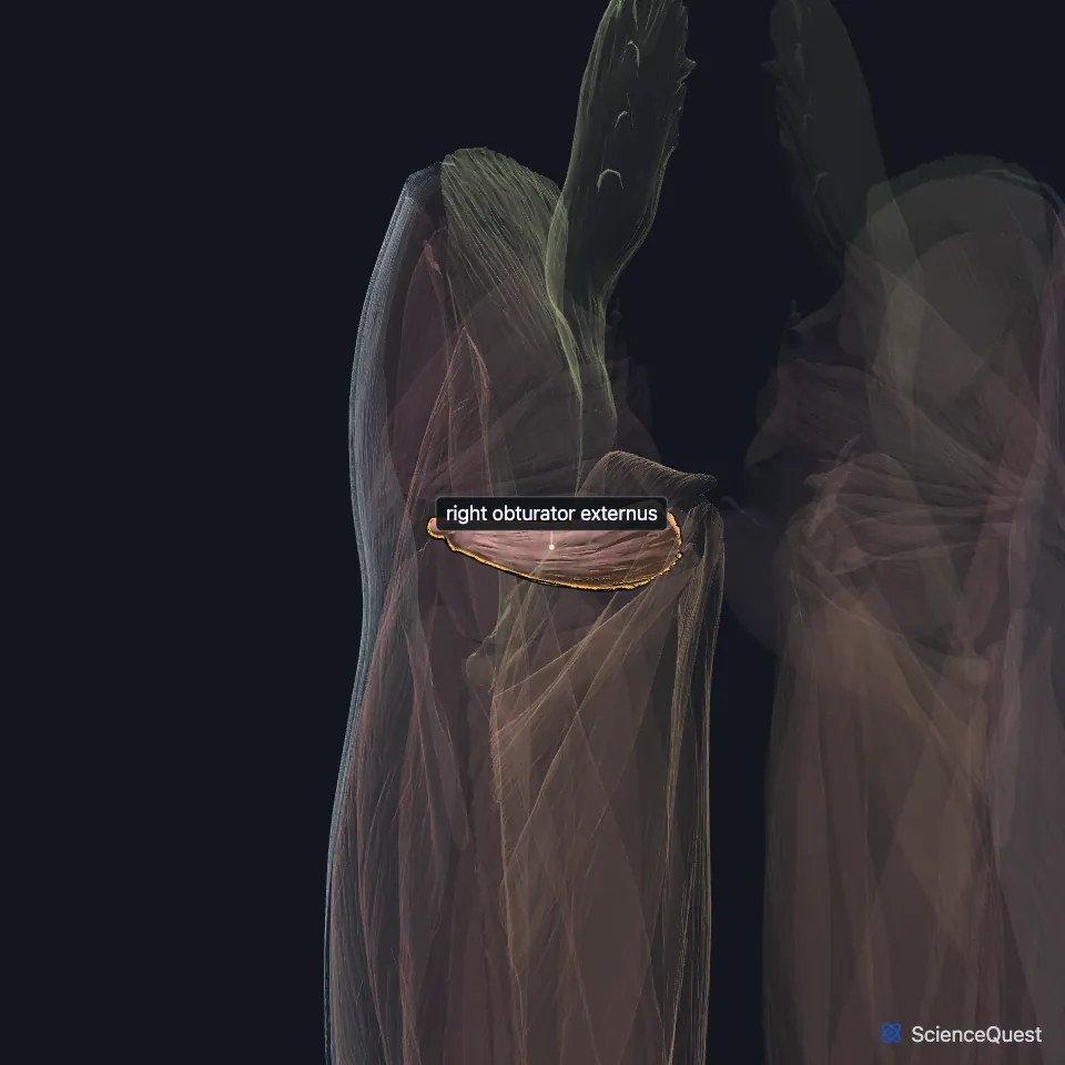
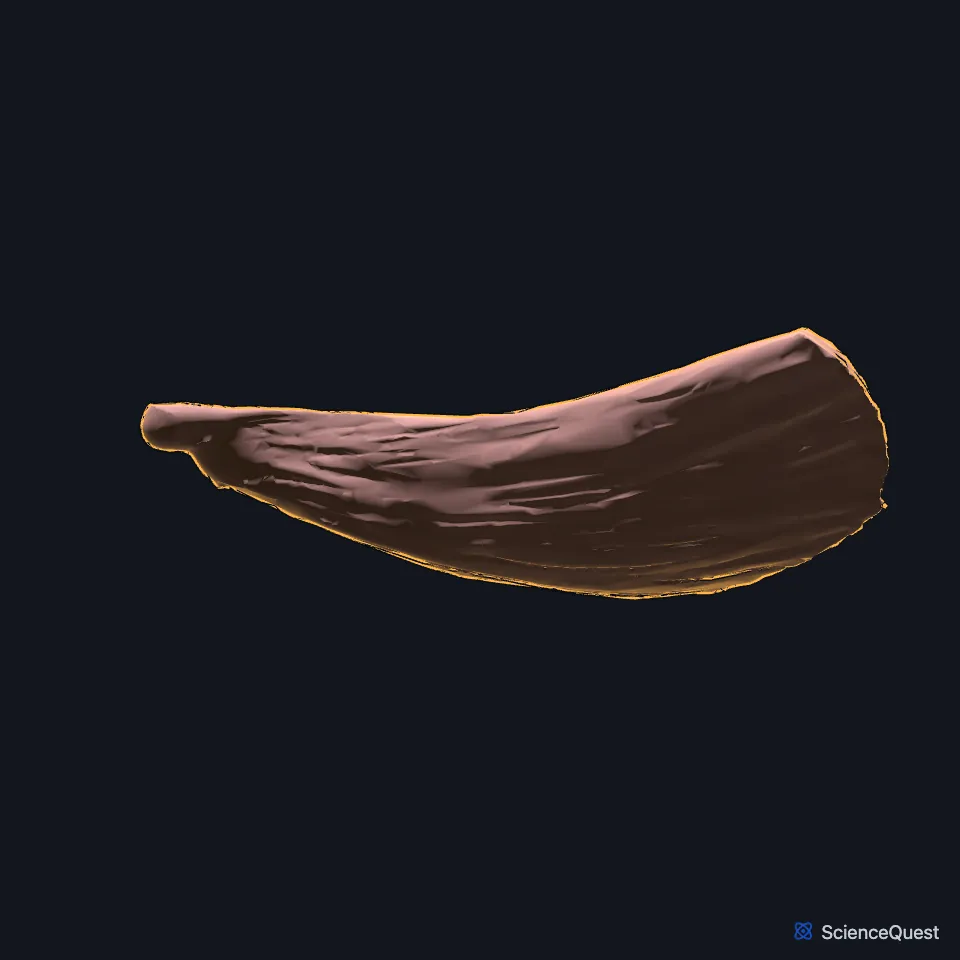
Open it in the Lower Limb Muscles Explorer See it alone in 3D
Where is the obturator externus?
It arises from the bone around the medial side of the obturator foramen, that is the rami of the pubis and the inferior ramus of the ischium, and from the medial two thirds of the outer surface of the obturator membrane. (Gray’s Anatomy (1918), Muscles of the Gluteal Region)
StatPearls notes that most of its fibers come from the bony margin, and only some from the membrane. (StatPearls, Obturator Muscles)
Where does the obturator externus insert?
Its fibers run backward, outward and upward into a tendon. The tendon crosses the back of the femoral neck and the lower hip joint capsule to reach the trochanteric fossa of the femur. (Gray’s Anatomy (1918), Muscles of the Gluteal Region; StatPearls, Hip)
In a cadaver study it turned to tendon at the level of the femoral neck, and its fibers at the lower edge of the acetabulum acted like a sling under the neck. StatPearls reads this as possible reinforcement of the hip joint. (StatPearls, Obturator Muscles)
What does the obturator externus do?
Its main action is lateral rotation of the hip, with the hip neutral or flexed. It can also help adduct the flexed hip. (StatPearls, Obturator Muscles)
Gray’s (1918) notes that when you sit, the other short rotators stop rotating and become abductors, but the obturator externus still rotates the femur. (Gray’s Anatomy (1918), Muscles of the Gluteal Region)
Which nerve and vessels lie by the obturator externus?
The posterior branch of the obturator nerve, from L2 to L4 of the lumbar plexus, supplies it. The obturator vessels lie between the muscle and the obturator membrane. (StatPearls, Obturator Muscles; Gray’s Anatomy (1918), Muscles of the Gluteal Region)
The deep branch of the medial circumflex femoral artery, the main supply of the femoral head, runs along its lower border and crosses behind its tendon. One study found the muscle protected that branch during hip dislocation in any direction. (StatPearls, Hip and Obturator Muscles)
Because the obturator nerve runs through the pelvis, it can be injured in gynecological surgery such as lymph node removal for cancer. That could weaken the obturator externus but not the obturator internus, which has its own nerve. (StatPearls, Obturator Muscles)
Where the sources differ
Sources disagree on how the obturator nerve passes it. Gray’s (1918) has the anterior branch passing in front of the muscle and the posterior branch piercing it; the StatPearls hip article has the posterior division running behind it. (Gray’s Anatomy (1918), Muscles of the Gluteal Region; StatPearls, Hip)
Sources give different spinal roots for its nerve. The StatPearls obturator muscles article gives L2 to L4, the roots of the obturator nerve; Gray’s (1918) has the third and fourth lumbar nerves supplying it through that nerve. (StatPearls, Obturator Muscles; Gray’s Anatomy (1918), Muscles of the Gluteal Region)
| Feature | Detail and source |
|---|---|
| Origin | Bony margin of the obturator foramen (pubic rami, inferior ramus of the ischium) and the outer surface of the obturator membrane (Gray’s Anatomy (1918), Muscles of the Gluteal Region; StatPearls, Hip) |
| Insertion | Trochanteric fossa of the femur (Gray’s Anatomy (1918), Muscles of the Gluteal Region; StatPearls, Hip) |
| Action | Rotates the hip laterally, neutral or flexed; helps adduct the flexed hip (StatPearls, Obturator Muscles) |
| Nerve | Posterior branch of the obturator nerve (L2 to L4) (StatPearls, Obturator Muscles) |
Touches in this model
Measured on the BodyParts3D meshes: surfaces within 2 mm of the obturator externus, nearest first.
- Right obturator internus in contact
- Right quadratus femoris in contact
- Right femur in contact
- Right hip bone in contact
- Skin in contact
- Tendinous arch of levator ani in contact
- Right puborectalis 0.5 mm away
Sources
Sources read
- Anatomy of the Human Body (Gray’s Anatomy), 20th US edition, 1918, Myology, The Muscles and Fasciae of the Thigh, 3. The Muscles of the Gluteal Region, pages 474 to 478; Internet Archive scan of the 1918 Lea and Febiger printing, terminology of 1918. Henry Gray; edited by Warren H. Lewis. Public domain. Read .
- StatPearls, Anatomy, Abdomen and Pelvis, Obturator Muscles. Maddy R. Larson; Weston Ryan. Last update . CC BY-NC-ND 4.0. Read .
- StatPearls, Anatomy, Bony Pelvis and Lower Limb, Hip. Roland Glenister; Sandeep Sharma. Last update . CC BY-NC-ND 4.0. Read .
The values are facts and free to use; this page’s selection and presentation are © 2026 ScienceQuest.