Adductor brevis
The adductor brevis is a short, roughly triangular adductor of the thigh. It lies behind the pectineus and the adductor longus. (Gray’s Anatomy (1918), Medial Femoral Muscles)
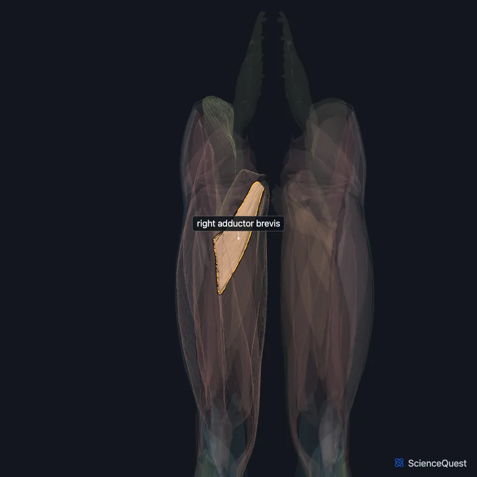
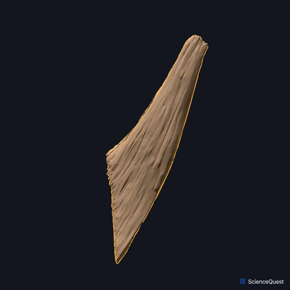
Open it in the Lower Limb Muscles Explorer See it alone in 3D
Where is the adductor brevis?
It arises by a narrow origin from the outer surfaces of the superior and inferior pubic rami, between the gracilis and the obturator externus. Its fibers run back, out and down to the femur. (Gray’s Anatomy (1918), Medial Femoral Muscles)
It inserts on the line from the lesser trochanter to the linea aspera and on the upper linea aspera, just behind the pectineus and the upper adductor longus. (Gray’s Anatomy (1918), Medial Femoral Muscles; StatPearls, Medial Thigh Muscles)
A small part of it may join the iliopsoas, pectineus and adductor longus in the floor of the femoral triangle. (StatPearls, Adductor Canal; Gray’s Anatomy (1918), The Femoral Artery)
What does the adductor brevis do?
It adducts the thigh and helps flex it at the hip. In walking, with the other adductors, it helps draw the lower limb forward. (StatPearls, Thigh Muscles; Gray’s Anatomy (1918), Medial Femoral Muscles)
How do the obturator nerve’s branches pass the adductor brevis?
The two branches of the obturator nerve lie on either side of it. The anterior branch runs between the adductor longus and the adductor brevis, and the posterior branch between the adductor brevis and the adductor magnus. (StatPearls, Obturator Nerve)
The obturator nerve supplies it. Its supply can come from the anterior branch, the posterior branch, or both. (StatPearls, Thigh Muscles and Obturator Nerve)
On MRI, wasting of the adductor brevis or the adductor longus can point to entrapment of the obturator nerve. (StatPearls, Medial Thigh Muscles)
How does the adductor brevis vary?
It may be divided into two or three parts, or it may be united with the adductor magnus. (Gray’s Anatomy (1918), Medial Femoral Muscles)
Where the sources differ
Sources disagree on how it rotates the thigh. The StatPearls thigh muscles article and Gray’s (1918) say it rotates the thigh laterally; a study the StatPearls adductor magnus article cites found a slight medial rotation lever arm in upright standing. (StatPearls, Thigh Muscles and Thigh Adductor Magnus Muscle; Gray’s Anatomy (1918), Medial Femoral Muscles)
Sources differ on its share of the femoral triangle’s floor. The StatPearls adductor canal article lists it among the floor muscles; Gray’s (1918) includes a small part of it, and only in some cases. (StatPearls, Adductor Canal; Gray’s Anatomy (1918), The Femoral Artery)
| Feature | Detail and source |
|---|---|
| Origin | Superior and inferior pubic rami (Gray’s); body and inferior pubic ramus (StatPearls) (Gray’s Anatomy (1918), Medial Femoral Muscles; StatPearls, Hip) |
| Insertion | Line from the lesser trochanter to the linea aspera, and the upper linea aspera (Gray’s Anatomy (1918), Medial Femoral Muscles) |
| Action | Adducts the thigh; helps flex it (StatPearls, Thigh Muscles) |
| Nerve | Obturator nerve, anterior branch, posterior branch or both (StatPearls, Thigh Muscles) |
Touches in this model
Measured on the BodyParts3D meshes: surfaces within 2 mm of the adductor brevis, nearest first.
- Right adductor longus in contact
- Right adductor magnus in contact
- Right adductor minimus in contact
- Right pectineus in contact
- Right vastus medialis in contact
- Right femur in contact
- Right hip bone in contact
- Skin in contact
Sources
Sources read between and
- Anatomy of the Human Body (Gray’s Anatomy), 20th US edition, 1918, Myology, The Muscles and Fasciae of the Thigh, 2. The Medial Femoral Muscles, pages 471 to 474; Internet Archive scan of the 1918 Lea and Febiger printing, terminology of 1918. Henry Gray; edited by Warren H. Lewis. Public domain. Read .
- StatPearls, Anatomy, Bony Pelvis and Lower Limb: Medial Thigh Muscles. Jordan L. Ramage; Matthew A. Varacallo. Last update . CC BY-NC-ND 4.0. Read .
- StatPearls, Anatomy, Abdomen and Pelvis: Adductor Canal (Subsartorial Canal, Hunter Canal). Allan Migirov; Tafline C. Arbor; Renato C. Vilella. Last update . CC BY-NC-ND 4.0. Read .
- Anatomy of the Human Body (Gray’s Anatomy), 20th US edition, 1918, Angiology, The Arteries of the Lower Extremity, The Femoral Artery, pages 626 to 627; Internet Archive scan of the 1918 Lea and Febiger printing, terminology of 1918. Henry Gray; edited by Warren H. Lewis. Public domain. Read .
- StatPearls, Anatomy, Bony Pelvis and Lower Limb: Thigh Muscles. Basem Attum; Matthew A. Varacallo. Last update . CC BY-NC-ND 4.0. Read .
- StatPearls, Anatomy, Abdomen and Pelvis, Obturator Nerve. Matthew Koh; Brian Markovich. Last update . CC BY-NC-ND 4.0. Read .
- StatPearls, Anatomy, Bony Pelvis and Lower Limb, Hip. Roland Glenister; Sandeep Sharma. Last update . CC BY-NC-ND 4.0. Read .
- StatPearls, Anatomy, Bony Pelvis and Lower Limb: Thigh Adductor Magnus Muscle. Susan H. Jeno; Marjorie V. Launico; Gary S. Schindler. Last update . CC BY-NC-ND 4.0. Read .
The values are facts and free to use; this page’s selection and presentation are © 2026 ScienceQuest.