Pectineus
The pectineus is a flat, four-sided muscle at the top of the inner thigh that flexes and adducts the hip. It counts as a transitional muscle between the front and medial thigh groups, because the femoral nerve, and sometimes the obturator nerve, supplies it. (StatPearls, Anterior Thigh Muscles and Hip)
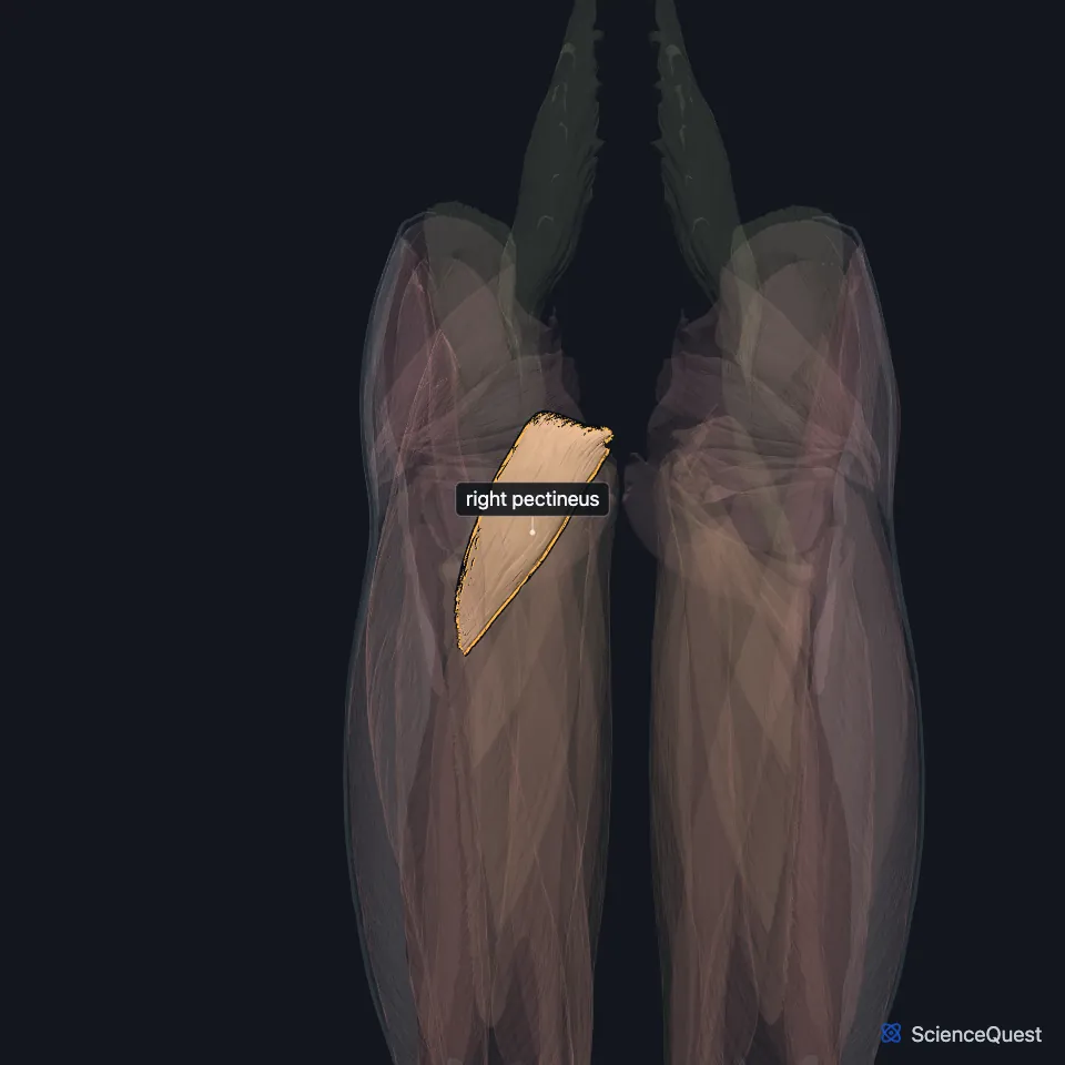
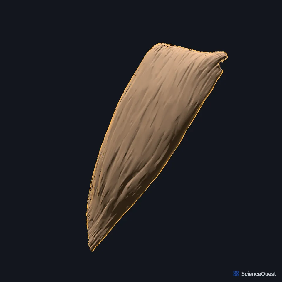
Open it in the Lower Limb Muscles Explorer See it alone in 3D
Where is the pectineus?
It arises from the pectineal line of the pubis and inserts on the back of the femur, along the line from the lesser trochanter to the linea aspera. (StatPearls, Medial Thigh Muscles; Gray’s Anatomy (1918), Medial Femoral Muscles)
With the iliopsoas and adductor longus, and sometimes a small part of the adductor brevis, it forms the floor of the femoral triangle. (StatPearls, Adductor Canal and Anterior Thigh Muscles; Gray’s Anatomy (1918), The Femoral Artery)
The adductor longus lies in the same plane, and the adductor brevis lies behind both. (Gray’s Anatomy (1918), Medial Femoral Muscles)
Where does the name pectineus come from?
Pecten is Latin for a comb. The muscle is named for the parallel fibers at its insertion, which look like the teeth of a comb. (StatPearls, Thigh Femoral Nerve)
What does the pectineus do?
It flexes and adducts the hip. (StatPearls, Hip and Thigh Femoral Nerve)
It assists the psoas major and iliacus, the main hip flexors. In walking, it helps draw the lower limb forward. (StatPearls, Hip Joint; Gray’s Anatomy (1918), Medial Femoral Muscles)
Which nerves supply the pectineus?
The femoral nerve supplies it through the nerve to the pectineus, a motor branch of the nerve’s anterior division. Sometimes the obturator nerve adds a branch. (StatPearls, Thigh Femoral Nerve and Anterior Thigh Muscles)
The deep branch of the medial circumflex femoral artery runs between the pectineus on its medial side and the iliopsoas tendon on its lateral side. (StatPearls, Hip)
How does the pectineus vary?
It may be split into an outer part supplied by the femoral nerve and an inner part supplied by the obturator nerve. A footnote in Gray’s (1918), quoting Paterson, calls the outer layer constant and has the accessory obturator nerve supply it when the femoral branch is missing. The muscle may attach to the hip joint capsule, and the adductor longus may be partly joined to it. (Gray’s Anatomy (1918), Medial Femoral Muscles)
Where the sources differ
Sources disagree on which way it rotates the thigh. The StatPearls anterior thigh article lists medial rotation; Gray’s (1918) says the pectineus and the adductors rotate the thigh laterally, because of the slant of their insertions. (StatPearls, Anterior Thigh Muscles; Gray’s Anatomy (1918), Medial Femoral Muscles)
| Feature | Detail and source |
|---|---|
| Origin | Pectineal line of the pubis (StatPearls, Hip; Gray’s Anatomy (1918), Medial Femoral Muscles) |
| Insertion | Back of the femur, on the line from the lesser trochanter to the linea aspera (StatPearls, Medial Thigh Muscles; Gray’s Anatomy (1918), Medial Femoral Muscles) |
| Action | Flexes and adducts the hip (StatPearls, Hip) |
| Nerve | Femoral nerve (L2 to L4); sometimes also the obturator nerve (Gray’s Anatomy (1918), Medial Femoral Muscles; StatPearls, Anterior Thigh Muscles) |
Touches in this model
Measured on the BodyParts3D meshes: surfaces within 2 mm of the pectineus, nearest first, 12 of 15.
- Right adductor brevis in contact
- Right adductor longus in contact
- Right adductor minimus in contact
- Right psoas major in contact
- Right rectus femoris in contact
- Right vastus medialis in contact
- Right deferent duct in contact
- Right femur in contact
- Right hip bone in contact
- Right inguinal ligament in contact
- Right pyramidalis in contact
- Right transversus abdominis in contact
Sources
Sources read between and
- StatPearls, Anatomy, Bony Pelvis and Lower Limb: Anterior Thigh Muscles. Ayesha Khan; Abdul Arain. Last update . CC BY-NC-ND 4.0. Read .
- StatPearls, Anatomy, Bony Pelvis and Lower Limb, Hip. Roland Glenister; Sandeep Sharma. Last update . CC BY-NC-ND 4.0. Read .
- StatPearls, Anatomy, Bony Pelvis and Lower Limb: Medial Thigh Muscles. Jordan L. Ramage; Matthew A. Varacallo. Last update . CC BY-NC-ND 4.0. Read .
- Anatomy of the Human Body (Gray’s Anatomy), 20th US edition, 1918, Myology, The Muscles and Fasciae of the Thigh, 2. The Medial Femoral Muscles, pages 471 to 474; Internet Archive scan of the 1918 Lea and Febiger printing, terminology of 1918. Henry Gray; edited by Warren H. Lewis. Public domain. Read .
- StatPearls, Anatomy, Abdomen and Pelvis: Adductor Canal (Subsartorial Canal, Hunter Canal). Allan Migirov; Tafline C. Arbor; Renato C. Vilella. Last update . CC BY-NC-ND 4.0. Read .
- Anatomy of the Human Body (Gray’s Anatomy), 20th US edition, 1918, Angiology, The Arteries of the Lower Extremity, The Femoral Artery, pages 626 to 627; Internet Archive scan of the 1918 Lea and Febiger printing, terminology of 1918. Henry Gray; edited by Warren H. Lewis. Public domain. Read .
- StatPearls, Anatomy, Bony Pelvis and Lower Limb: Thigh Femoral Nerve. Nader A. Refai; Asa C. Black; Prasanna Tadi. Last update . CC BY-NC-ND 4.0. Read .
- StatPearls, Anatomy, Bony Pelvis and Lower Limb, Hip Joint. Maks Gold; Akul Munjal; Matthew A. Varacallo. Last update . CC BY-NC-ND 4.0. Read .
The values are facts and free to use; this page’s selection and presentation are © 2026 ScienceQuest.