Gracilis
The gracilis is a long, thin muscle on the inner side of the thigh, the most superficial of the medial thigh group. Unlike the other adductors, it attaches to the tibia rather than the femur. (StatPearls, Thigh Gracilis Muscle and Thigh Adductor Magnus Muscle; Gray’s Anatomy (1918), Medial Femoral Muscles)
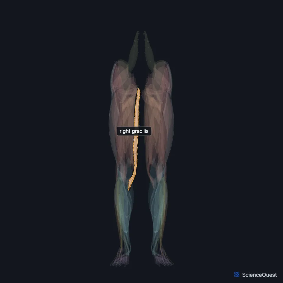
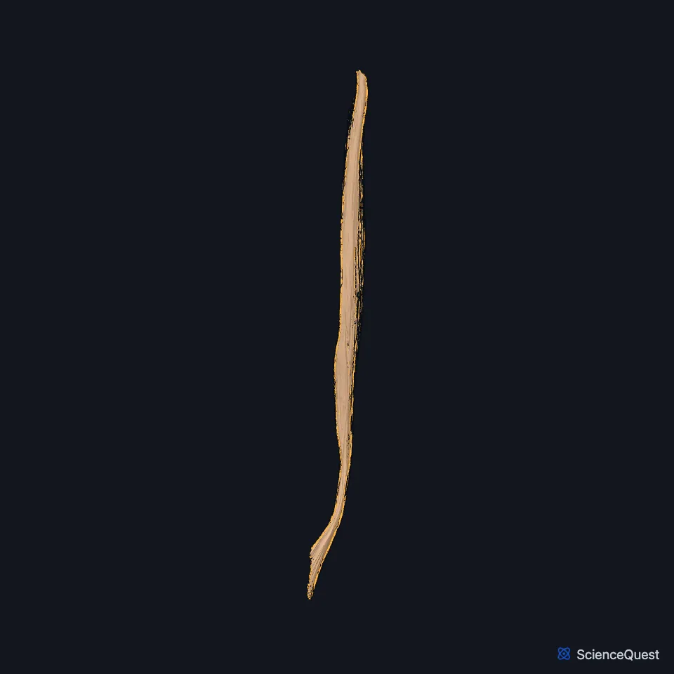
Open it in the Lower Limb Muscles Explorer See it alone in 3D
Where is the gracilis?
It is thin and flat, broad above and tapering below. It arises by a thin aponeurosis from the lower half of the pubic symphysis and the upper half of the pubic arch. (Gray’s Anatomy (1918), Medial Femoral Muscles)
Its rounded tendon passes behind the medial condyle of the femur, curves around the medial condyle of the tibia, and inserts on the upper medial surface of the tibia. (Gray’s Anatomy (1918), Medial Femoral Muscles)
What is the pes anserinus?
At the tibia its tendon joins those of the sartorius and semitendinosus in the pes anserinus, named for its likeness to a goose’s foot. (StatPearls, Medial Thigh Muscles and Thigh Gracilis Muscle)
Its tendon lies just above the semitendinosus tendon, and the sartorius tendon overlaps its upper edge. A bursa it shares with the semitendinosus separates it from the tibial collateral ligament. (Gray’s Anatomy (1918), Medial Femoral Muscles)
Its fiber bundles spiral onto the front of its tendon, which makes it a spiral unipennate muscle. They form 5 to 7 compartments, each with its own nerve branches, which may work independently. (StatPearls, Thigh Gracilis Muscle)
What does the gracilis do?
It crosses both the hip and the knee. At the hip it adducts, flexes and rotates medially; at the knee it flexes and rotates the leg medially. (StatPearls, Thigh Gracilis Muscle; Gray’s Anatomy (1918), Medial Femoral Muscles)
Surgeons can use its tendon to rebuild the anterior cruciate ligament. The muscle can also serve as a free flap, because its nerve and vessels enter at predictable pedicles. (StatPearls, Thigh Gracilis Muscle)
Which nerve and neighbors does the gracilis have?
The anterior branch of the obturator nerve supplies it. The nerve enters with the main artery and two veins at a major pedicle, usually one, on the upper front of the muscle. (StatPearls, Thigh Gracilis Muscle)
The saphenous nerve leaves the adductor canal by passing between the gracilis and the sartorius, then runs on to the skin of the medial leg. (StatPearls, Medial Thigh Muscles)
Pain where its tendon becomes the pes anserinus, especially with swelling, suggests pes anserinus bursitis. That condition can mimic an injury inside the knee such as a meniscal tear. (StatPearls, Thigh Gracilis Muscle)
Where the sources differ
Sources word its origin differently. StatPearls gives the medial ischiopubic ramus in its gracilis article and the body and inferior ramus of the pubis in its medial thigh article; Gray’s (1918) gives the lower half of the symphysis and the upper half of the pubic arch. (StatPearls, Thigh Gracilis Muscle and Medial Thigh Muscles; Gray’s Anatomy (1918), Medial Femoral Muscles)
| Feature | Detail and source |
|---|---|
| Origin | Lower half of the pubic symphysis and upper half of the pubic arch (Gray’s); body and inferior ramus of the pubis, or the ischiopubic ramus (StatPearls) (Gray’s Anatomy (1918), Medial Femoral Muscles; StatPearls, Medial Thigh Muscles and Thigh Gracilis Muscle) |
| Insertion | Upper medial surface of the tibia, in the pes anserinus (StatPearls, Thigh Gracilis Muscle) |
| Action | Adducts, flexes and medially rotates the hip; flexes the knee and rotates it medially (StatPearls, Thigh Gracilis Muscle) |
| Nerve | Anterior branch of the obturator nerve (StatPearls, Thigh Gracilis Muscle) |
Touches in this model
Measured on the BodyParts3D meshes: surfaces within 2 mm of the gracilis, nearest first.
- Right adductor longus in contact
- Right adductor magnus in contact
- Right adductor minimus in contact
- Right sartorius in contact
- Right semimembranosus in contact
- Corpus cavernosum of penis in contact
- Right hip bone in contact
- Skin in contact
- Right semitendinosus 0.4 mm away
Sources
Sources read
- StatPearls, Anatomy, Bony Pelvis and Lower Limb: Thigh Gracilis Muscle. Irfan A. Khan; Bruno Bordoni; Matthew A. Varacallo. Last update . CC BY-NC-ND 4.0. Read .
- StatPearls, Anatomy, Bony Pelvis and Lower Limb: Thigh Adductor Magnus Muscle. Susan H. Jeno; Marjorie V. Launico; Gary S. Schindler. Last update . CC BY-NC-ND 4.0. Read .
- Anatomy of the Human Body (Gray’s Anatomy), 20th US edition, 1918, Myology, The Muscles and Fasciae of the Thigh, 2. The Medial Femoral Muscles, pages 471 to 474; Internet Archive scan of the 1918 Lea and Febiger printing, terminology of 1918. Henry Gray; edited by Warren H. Lewis. Public domain. Read .
- StatPearls, Anatomy, Bony Pelvis and Lower Limb: Medial Thigh Muscles. Jordan L. Ramage; Matthew A. Varacallo. Last update . CC BY-NC-ND 4.0. Read .
The values are facts and free to use; this page’s selection and presentation are © 2026 ScienceQuest.