Semimembranosus
The semimembranosus is one of the three hamstring muscles, at the back and inner side of the thigh. It is named for its membranous tendon of origin. (Gray’s Anatomy (1918), Posterior Femoral Muscles; StatPearls, Hamstring Muscle)
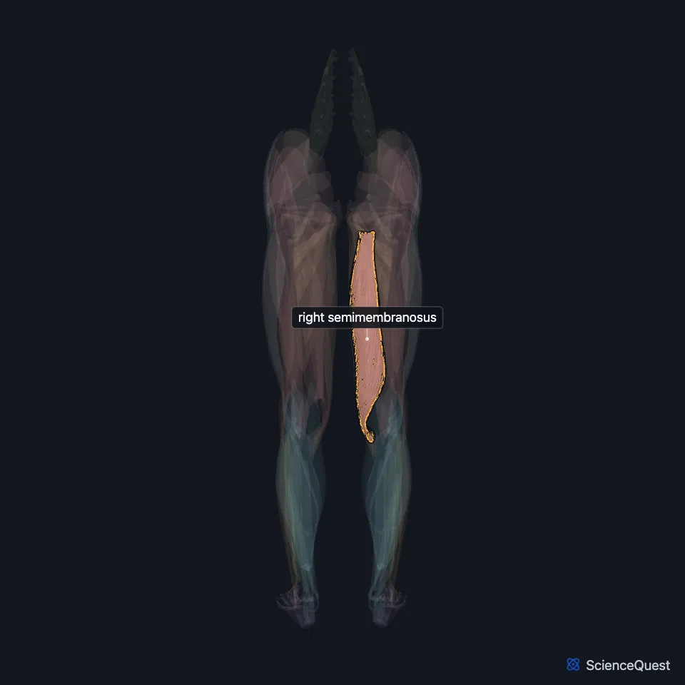
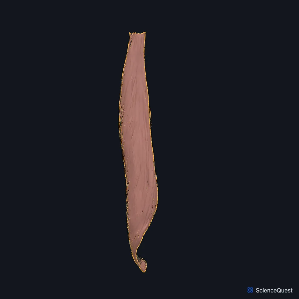
Open it in the Lower Limb Muscles Explorer See it alone in 3D
Where is the semimembranosus?
It arises by a thick tendon from the upper outer part of the ischial tuberosity, above and lateral to the biceps femoris and semitendinosus. It lies deep to the semitendinosus. (Gray’s Anatomy (1918), Posterior Femoral Muscles; StatPearls, Posterior Thigh)
Above the knee, with the semitendinosus, it forms the upper medial border of the popliteal fossa. It overlaps the upper part of the popliteal vessels. (StatPearls, Posterior Thigh Muscles; Gray’s Anatomy (1918), Posterior Femoral Muscles)
Where does the semimembranosus insert?
Its tendon of origin spreads into an aponeurosis over the upper front of the muscle. The fibers run to a second aponeurosis on the lower back of the muscle, which narrows into the tendon of insertion. (Gray’s Anatomy (1918), Posterior Femoral Muscles)
It inserts mainly into the horizontal groove on the back of the medial condyle of the tibia. (Gray’s Anatomy (1918), Posterior Femoral Muscles; StatPearls, Posterior Thigh Muscles)
The tendon sends out expansions. A large one runs up and out to the back of the lateral condyle of the femur, forming part of the oblique popliteal ligament, and others reach the fascia over the popliteus and the tibial collateral ligament. (Gray’s Anatomy (1918), Posterior Femoral Muscles)
In the study StatPearls cites, it averaged 38.7 cm long, shorter than the semitendinosus at 44.3 cm. (StatPearls, Hamstring Muscle)
What does the semimembranosus do?
It extends the hip and flexes the knee. With the knee bent, it also rotates the tibia medially. (StatPearls, Posterior Thigh Muscles and Hamstring Muscle)
With the other hamstrings it helps raise the trunk from a stoop. The hamstrings are short enough that the hip cannot flex fully unless the knee bends too. (Gray’s Anatomy (1918), Posterior Femoral Muscles)
A type 2 hamstring strain, from overstretching, most often with the hip flexed and the knee straight, injures the proximal tendon of the semimembranosus. It takes longer to heal than the type 1 strain of the biceps femoris. (StatPearls, Posterior Thigh Muscles)
Which nerve and vessels supply the semimembranosus?
The tibial nerve, the tibial part of the sciatic nerve, supplies it, usually by a single branch. (StatPearls, Hamstring Muscle and Posterior Thigh Muscles)
Its blood comes from the perforating branches of the profunda femoris artery, the inferior gluteal artery and upper muscular branches of the popliteal artery. (StatPearls, Posterior Thigh Muscles)
Can the semimembranosus be missing?
It may be small, absent or doubled. StatPearls reports a case where both semimembranosus muscles were missing, found by chance on MRI after a fall. (Gray’s Anatomy (1918), Posterior Femoral Muscles; StatPearls, Hamstring Muscle)
| Feature | Detail and source |
|---|---|
| Origin | Upper outer part of the ischial tuberosity (StatPearls, Posterior Thigh Muscles; Gray’s Anatomy (1918), Posterior Femoral Muscles) |
| Insertion | Groove on the back of the medial condyle of the tibia (StatPearls, Posterior Thigh Muscles; Gray’s Anatomy (1918), Posterior Femoral Muscles) |
| Action | Extends the hip; flexes the knee; rotates the tibia medially with the knee bent (StatPearls, Posterior Thigh Muscles) |
| Nerve | Tibial nerve, from the sciatic nerve (StatPearls, Posterior Thigh Muscles) |
Touches in this model
Measured on the BodyParts3D meshes: surfaces within 2 mm of the semimembranosus, nearest first.
- Long head of right biceps femoris in contact
- Medial head of right gastrocnemius in contact
- Right adductor magnus in contact
- Right gracilis in contact
- Right semitendinosus in contact
- Right femur in contact
- Right hip bone in contact
- Right tibia in contact
- Skin in contact
- Right sartorius 1.3 mm away
- Corpus cavernosum of penis 1.3 mm away
Sources
Sources read
- Anatomy of the Human Body (Gray’s Anatomy), 20th US edition, 1918, Myology, The Muscles and Fasciae of the Thigh, 4. The Posterior Femoral Muscles (Hamstring Muscles), pages 478 to 479; Internet Archive scan of the 1918 Lea and Febiger printing, terminology of 1918. Henry Gray; edited by Warren H. Lewis. Public domain. Read .
- StatPearls, Anatomy, Bony Pelvis and Lower Limb, Hamstring Muscle. Young Hu; Avais Raja. Last update . CC BY-NC-ND 4.0. Read .
- StatPearls, Anatomy, Bony Pelvis and Lower Limb: Posterior Thigh. Thomas B. Anderson; Renato C. Vilella. Last update . CC BY-NC-ND 4.0. Read .
- StatPearls, Anatomy, Bony Pelvis and Lower Limb: Posterior Thigh Muscles. Jacob E. Vaughn; Wayne B. Cohen-Levy. Last update . CC BY-NC-ND 4.0. Read .
The values are facts and free to use; this page’s selection and presentation are © 2026 ScienceQuest.