Adductor magnus
The adductor magnus is the largest and most posterior muscle of the medial thigh. Its adductor and hamstring parts each have their own distal attachment, and the two parts usually get different nerves. (StatPearls, Thigh Adductor Magnus Muscle)
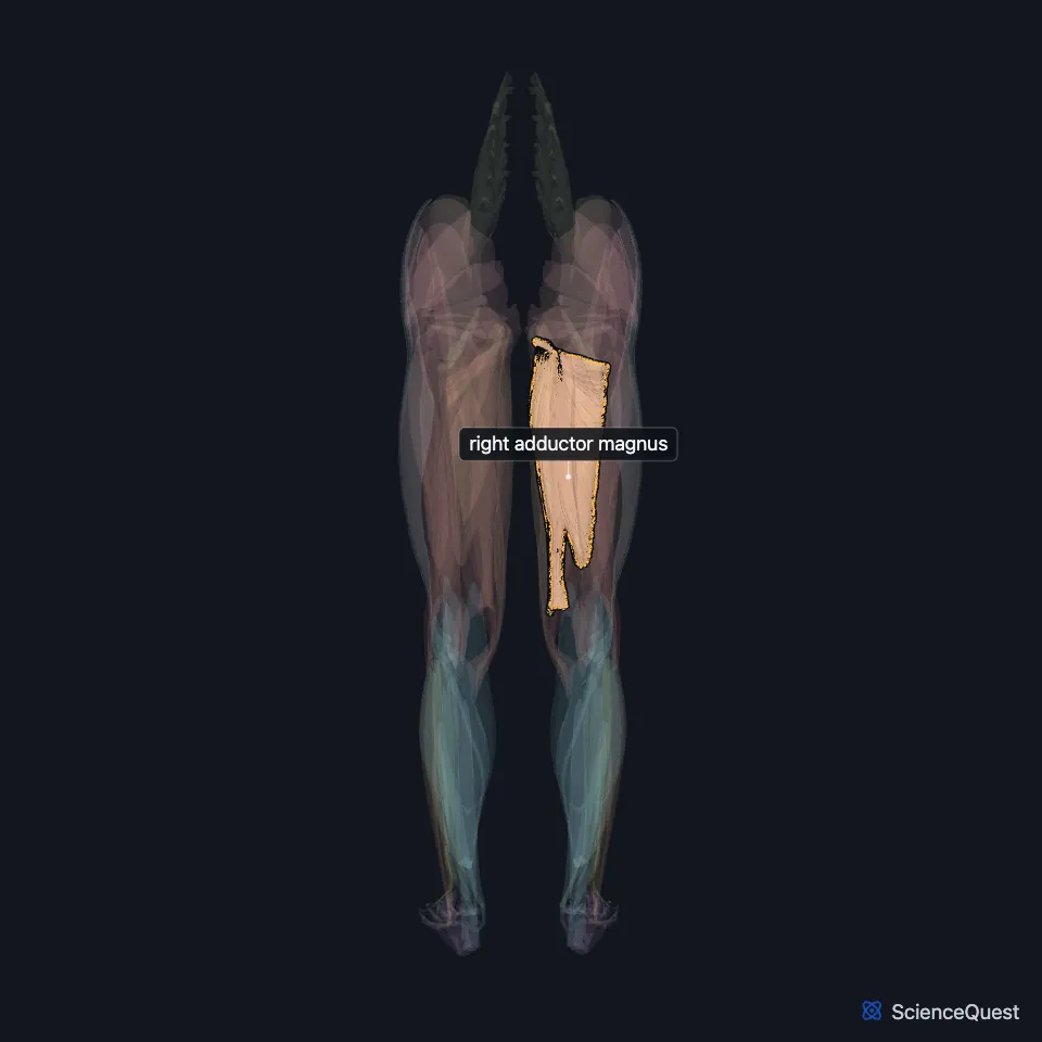
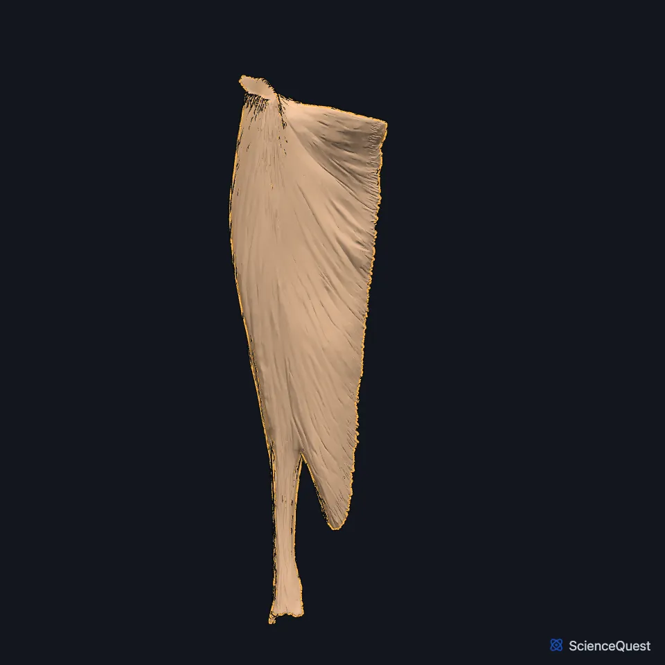
Open it in the Lower Limb Muscles Explorer See it alone in 3D
Where is the adductor magnus?
It is a large triangular muscle on the medial side of the thigh. It arises from the inferior pubic ramus, the ischial ramus and the ischial tuberosity. (StatPearls, Thigh Adductor Magnus Muscle; Gray’s Anatomy (1918), Medial Femoral Muscles)
At the back of the thigh, the sciatic nerve runs down on its surface, deep to the hamstrings and the long head of the biceps femoris. (StatPearls, Hip and Sciatic Nerve)
What are the two parts of the adductor magnus?
The adductor part runs from the pubic and ischial rami to the linea aspera and the upper medial supracondylar line. Its short, more horizontal upper fibers reach the gluteal tuberosity, and some authors call them the adductor minimus. (StatPearls, Thigh Adductor Magnus Muscle)
The hamstring part runs almost vertically from the ischial tuberosity to the adductor tubercle on the medial condyle of the femur. The adductor part makes up about 70% of the muscle. (StatPearls, Thigh Adductor Magnus Muscle)
Near its insertion are five openings framed by tendinous arches. The upper four carry the perforating branches of the profunda femoris artery, and the largest, the adductor hiatus, lets the femoral vessels pass from the adductor canal to the popliteal fossa. (StatPearls, Thigh Adductor Magnus Muscle; Gray’s Anatomy (1918), Medial Femoral Muscles)
What does the adductor magnus do?
Both parts adduct the thigh and steady the pelvis during walking. The adductor part also flexes the thigh, and the hamstring part also extends it. (StatPearls, Thigh Adductor Magnus Muscle)
StatPearls compares it to the deltoid. One part flexes and medially rotates the thigh, another extends and laterally rotates it, and both stabilize a ball-and-socket joint. (StatPearls, Thigh Adductor Magnus Muscle)
Which nerves and muscles work with the adductor magnus?
It has two nerves. The posterior division of the obturator nerve (L2 to L4) supplies the adductor part, and the tibial part of the sciatic nerve supplies the hamstring part. (StatPearls, Thigh Adductor Magnus Muscle; Gray’s Anatomy (1918), Medial Femoral Muscles)
Its insertion on the linea aspera merges with the origin of the short head of the biceps femoris, and the two work together to stabilize the femur and pelvis. (StatPearls, Thigh Adductor Magnus Muscle)
Its tendon sits medial to the hamstring origin on the ischial tuberosity. On MRI an intact adductor magnus can mask a complete hamstring avulsion, looking like a thinned but intact semimembranosus tendon. (StatPearls, Thigh Adductor Magnus Muscle)
How does the adductor magnus vary?
Its upper part is often described as a separate muscle, the adductor minimus. In one cadaver study, more than half of 20 adults and all 5 fetuses had an adductor minimus distinct from the adductor magnus. (Gray’s Anatomy (1918), Medial Femoral Muscles; StatPearls, Thigh Adductor Magnus Muscle)
It may be fused with the quadratus femoris. (Gray’s Anatomy (1918), Medial Femoral Muscles)
Where the sources differ
Sources disagree on how cleanly the two nerves divide the muscle. The usual account gives the adductor part to the obturator nerve and the hamstring part to the sciatic nerve; Takizawa and colleagues, cited by StatPearls, found both nerves in the adductor part, with dual supply in over 90% of its lower segment. (StatPearls, Thigh Adductor Magnus Muscle and Sciatic Nerve)
Sources give different spinal roots for its obturator supply. The StatPearls adductor magnus article gives L2 to L4 for the posterior division that reaches the adductor part; Gray’s (1918) has the third and fourth lumbar nerves supplying all three adductors through the obturator nerve. (StatPearls, Thigh Adductor Magnus Muscle; Gray’s Anatomy (1918), Medial Femoral Muscles)
| Feature | Detail and source |
|---|---|
| Origin | Inferior pubic ramus, ischial ramus and ischial tuberosity (StatPearls, Thigh Adductor Magnus Muscle) |
| Insertion | Gluteal tuberosity, linea aspera and medial supracondylar line (adductor part); adductor tubercle of the femur (hamstring part) (StatPearls, Thigh Adductor Magnus Muscle) |
| Action | Adducts the thigh; the adductor part flexes it, the hamstring part extends it (StatPearls, Thigh Adductor Magnus Muscle) |
| Nerve | Obturator nerve, posterior division (adductor part); tibial part of the sciatic nerve (hamstring part) (StatPearls, Thigh Adductor Magnus Muscle) |
Touches in this model
Measured on the BodyParts3D meshes: surfaces within 2 mm of the adductor magnus, nearest first, 12 of 16.
- Long head of right biceps femoris in contact
- Medial head of right gastrocnemius in contact
- Right adductor brevis in contact
- Right adductor longus in contact
- Right adductor minimus in contact
- Right gracilis in contact
- Right psoas major in contact
- Right semimembranosus in contact
- Right vastus medialis in contact
- Short head of right biceps femoris in contact
- Right femur in contact
- Right hip bone in contact
Sources
Sources read
- StatPearls, Anatomy, Bony Pelvis and Lower Limb: Thigh Adductor Magnus Muscle. Susan H. Jeno; Marjorie V. Launico; Gary S. Schindler. Last update . CC BY-NC-ND 4.0. Read .
- Anatomy of the Human Body (Gray’s Anatomy), 20th US edition, 1918, Myology, The Muscles and Fasciae of the Thigh, 2. The Medial Femoral Muscles, pages 471 to 474; Internet Archive scan of the 1918 Lea and Febiger printing, terminology of 1918. Henry Gray; edited by Warren H. Lewis. Public domain. Read .
- StatPearls, Anatomy, Bony Pelvis and Lower Limb, Hip. Roland Glenister; Sandeep Sharma. Last update . CC BY-NC-ND 4.0. Read .
- StatPearls, Anatomy, Sciatic Nerve. Brittney A. Giuffre; Asa C. Black; Rebecca Jeanmonod. Last update . CC BY-NC-ND 4.0. Read .
The values are facts and free to use; this page’s selection and presentation are © 2026 ScienceQuest.