Obturator internus
The obturator internus is a triangular muscle that lies partly inside the lesser pelvis and partly behind the hip joint. It is one of the short lateral rotators of the hip. (StatPearls, Obturator Muscles; Gray’s Anatomy (1918), Muscles of the Gluteal Region)
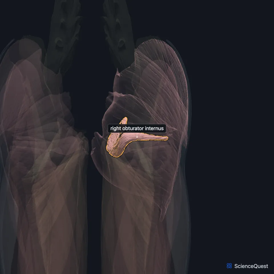
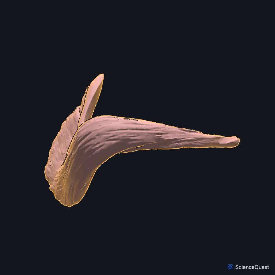
Open it in the Lower Limb Muscles Explorer See it alone in 3D
Where is the obturator internus?
Its fleshy part lines the inner surface of the anterolateral wall of the pelvis, around most of the obturator foramen. It arises from the pelvic surface of the obturator membrane and from the surrounding rami of the pubis and ischium. (Gray’s Anatomy (1918), Muscles of the Gluteal Region; StatPearls, Obturator Muscles)
The name comes from the Latin obturo, “to stop or block up”, because the obturator muscles cover the opening of the obturator foramen. (StatPearls, Obturator Muscles)
How does the obturator internus reach the femur?
Its fibers converge on the lesser sciatic foramen and end in four or five tendinous bands. These turn at a right angle around the grooved ischium between its spine and tuberosity, where a bursa separates tendon from bone. (Gray’s Anatomy (1918), Muscles of the Gluteal Region)
Outside the pelvis the bands join into one flat tendon that crosses the back of the hip joint capsule. The gemellus superior and gemellus inferior join it, and it inserts on the medial surface of the greater trochanter, above the trochanteric fossa. (Gray’s Anatomy (1918), Muscles of the Gluteal Region; StatPearls, Gemelli Muscles)
What does the obturator internus do?
It rotates the femur laterally when the hip is extended and abducts it when the hip is flexed. (StatPearls, Obturator Muscles; Gray’s Anatomy (1918), Muscles of the Gluteal Region)
It also holds the femoral head in the acetabulum. It shortens as the hip moves from flexion to extension, which suggests it supports the joint during weight-bearing and push-off. (StatPearls, Obturator Muscles)
Which nerve and vessels supply the obturator internus?
The nerve to the obturator internus, a branch of the sacral plexus, supplies it and the gemellus superior. The nerve leaves the pelvis through the greater sciatic foramen below the piriformis and comes back in through the lesser sciatic foramen to reach the muscle. (StatPearls, Obturator Muscles and Gemelli Muscles)
Branches of the obturator, inferior gluteal and pudendal arteries supply it. (StatPearls, Obturator Muscles)
The sciatic nerve runs deep to the piriformis and superficial to the obturator internus and the gemelli. Stretching this complex can compress the nerve, a condition called gemelli-obturator syndrome. (StatPearls, Gemelli Muscles)
In the posterior approach to the hip, surgeons first find the sciatic nerve and then detach the short lateral rotators, including the obturator internus. Repairing them at closure lowered posterior dislocation rates from 4.5% to 0.5% in the studies StatPearls cites. (StatPearls, Obturator Muscles)
Where the sources differ
Sources give different spinal roots for its nerve. StatPearls gives L5 to S2 in its obturator and hip articles, L4 to S2 in its gemelli article, and L5 and S1 in its femur article; Gray’s (1918) gives the first, second and third sacral nerves. (StatPearls, Obturator Muscles, Hip, Gemelli Muscles and Femur; Gray’s Anatomy (1918), Muscles of the Gluteal Region)
| Feature | Detail and source |
|---|---|
| Origin | Pelvic surface of the obturator membrane and the surrounding rami of the pubis and ischium (StatPearls, Obturator Muscles; Gray’s Anatomy (1918), Muscles of the Gluteal Region) |
| Insertion | Medial surface of the greater trochanter, with the gemelli (Gray’s Anatomy (1918), Muscles of the Gluteal Region; StatPearls, Hip) |
| Action | Rotates the femur laterally with the hip extended; abducts it with the hip flexed; holds the femoral head in the acetabulum (StatPearls, Obturator Muscles) |
| Nerve | Nerve to the obturator internus, from the sacral plexus (StatPearls, Obturator Muscles) |
Touches in this model
Measured on the BodyParts3D meshes: surfaces within 2 mm of the obturator internus, nearest first.
- Right gemellus inferior in contact
- Right gemellus superior in contact
- Right obturator externus in contact
- Right femur in contact
- Right hip bone in contact
- Right iliococcygeus in contact
- Right pubococcygeus in contact
- Right puborectalis in contact
- Skin in contact
- Tendinous arch of levator ani in contact
Sources
Sources read
- StatPearls, Anatomy, Abdomen and Pelvis, Obturator Muscles. Maddy R. Larson; Weston Ryan. Last update . CC BY-NC-ND 4.0. Read .
- Anatomy of the Human Body (Gray’s Anatomy), 20th US edition, 1918, Myology, The Muscles and Fasciae of the Thigh, 3. The Muscles of the Gluteal Region, pages 474 to 478; Internet Archive scan of the 1918 Lea and Febiger printing, terminology of 1918. Henry Gray; edited by Warren H. Lewis. Public domain. Read .
- StatPearls, Anatomy, Bony Pelvis and Lower Limb, Gemelli Muscles. Bradley Lezak; Dustin H. Massel. Last update . CC BY-NC-ND 4.0. Read .
- StatPearls, Anatomy, Bony Pelvis and Lower Limb, Hip. Roland Glenister; Sandeep Sharma. Last update . CC BY-NC-ND 4.0. Read .
- StatPearls, Anatomy, Bony Pelvis and Lower Limb: Femur. Amy Chang; Grant Breeland; Asa C. Black; John B. Hubbard. Last update . CC BY-NC-ND 4.0. Read .
The values are facts and free to use; this page’s selection and presentation are © 2026 ScienceQuest.